Contribute to better healthcare by designing and developing medical innovations for improved diagnostics, treatment, rehabilitation, prevention and a better quality of life.
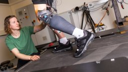
Can you think of friendlier, less painful or less harmful methods to detect breast cancer, or to perform an endoscopy? Can you pave the way for animal-free drug testing by developing mini organ-on-a-chip models, that can mimic an actual human organ, like a heart or liver? And what about detecting complex diseases like Parkinson’s or Alzheimer’s at an early stage, or developing an exoskeleton to train paralysed patients to walk? Advances in technologies are at the heart of innovation within healthcare. Are you eager to develop medical innovations that contribute to better care? If so, the Master’s in Biomedical Engineering at the University of Twente might just be right for you!
In this two-year, English-taught Master’s, you will learn to research, design, and develop innovative products and processes that will benefit the healthcare sector. With your expertise, you can contribute to the improvement of diagnostics, treatment and rehabilitation, but also to prevention and better quality of life. You will combine engineering skills in disciplines such as chemistry, physics, nanotechnology, electrical engineering and/or mechanical engineering with in-depth knowledge of biology and medicine. As a biomedical engineer, you can bridge the gap between healthcare and engineering, as you understand both contexts, thanks to the interdisciplinary character of this Master’s.
Start date
You can start your studies in September or February. However, the specialisation Bioengineering Technologies only starts in September. In case you need to follow the Pre-Master’s first, you can start in September and continue with the Master’s afterwards.
Choose a specialisation
You have a lot of freedom to tailor your Master’s in Biomedical Engineering to your interests and ambitions. Do you want to become an expert in medical imaging, or in measuring brain signals? Or do you want to contribute to the development of bionic prostheses or the creation of artificial (mini) organs from biomaterials? You will build your own expertise within one of the specialisations. Your choice determines which courses you will follow and the type of research you will engage in during your master’s thesis. You can choose from five different specialisations:
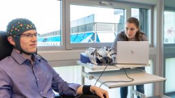
Innovate in the TechMed Centre
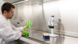
As a student in Biomedical Engineering, you will have unique access to state-of-the-art labs and other medical facilities on our high-tech campus. The programme is closely associated with UT’s renowned TechMed Centre, a leading institute that aims to improve healthcare through personalised technology. You have the opportunity to work with leading scientists and other experts from industry and clinical practice. It is a great environment for network building and gaining hands-on experience for your future career!
Career perspectives
Due to our ageing society, not only the number of patients seeking help for their health problems is increasing, but the health problems they report are also more complex. On many levels, healthcare is moving towards more automated processes in order to keep care efficient, effective, and more advanced (think of e.g. computer-aided diagnostics). In addressing this and virtually any other global health challenge, innovations in biomedical technologies are indispensable. As a biomedical engineer, you will be a key player in developing and advancing medical devices and clinical services, making you a highly sought-after professional on the job market. That’s why graduates of this Master’s find a job soon after graduation.
Programme overview
Related programmes
Below you find a list of educational programmes which are (closely) related to Biomedical Engineering.
Engineering & Technology
Applied Mathematics
Expand your understanding of mathematics to tackle challenging issues in a variety of sectors. This Master’s teaches you to discover new patterns and predict them through mathematical models.
Applied Physics
Deepen your understanding of the laws of physics to find technical solutions in a wide range of areas such as IT, sustainability, healthcare, chiptechnology, digitalisation, finance, space and more.
Business Information Technology
Become an expert in IT-based business innovation, able to devise new services and radically improve the way businesses work.
Chemical Science & Engineering
Develop new chemical processes, materials and molecules and contribute to innovations that benefit our society in areas such as health, water, food and sustainability.
Civil Engineering & Management
Do you want to work on future-proof solutions in the construction, water and/or transport sector? Learn to manage civil engineering projects with an eye for both technical and non-technical aspects.
Computer Science
Become an expert computer scientist and learn to solve the biggest challenges of our digital society by designing and developing more secure and efficient ICT systems.
Construction Management & Engineering
Become an expert in managing large-scale, complex construction projects in an increasingly digitised industry, using an innovative, integral approach.
Electrical Engineering
Design smart and sustainable electronics and advanced chips for applications such as biomedical technology, autonomous vehicles, computer vision, nanotechnology and the energy transition.
Embedded Systems
Become an expert in designing the software and hardware of complex embedded systems in vehicles, pacemakers and chipsets, amongst others.
Humanitarian Engineering
Do you want to tackle complex humanitarian crises worldwide? This Master’s helps you develop sustainable, socio-technological solutions to challenges faced by underserved and marginalised communities.
Industrial Design Engineering
Do you want to design innovative products that improve daily life and address today’s challenges? In this Master’s, you combine design and engineering to enhance both products and product development.
Industrial Engineering & Management
Combine technical expertise with management skills to analyse, improve, and future-proof complex business processes in (international) organisations.
Interaction Technology
Delve into the field of human-computer interaction and design and develop intelligent and interactive technologies that are meaningful to people and society.
Mechanical Engineering
Become an expert mechanical engineer who focuses on the futureproof design, analysis and maintenance of machinery, structures, products and production processes.
Nanotechnology
Tackle big challenges on the tiniest scale: become an expert in nanotechnology and develop smart, innovative solutions for major societal issues in healthcare, ICT and sustainability.
Philosophy of Science, Technology & Society
How does technology shape our lives and the world we live in? Learn to understand the role of modern technology in our society from a philosophical perspective.
Robotics
Become an expert in robotics: design smart, human-centred robots and combine AI, mechatronics, systems integration and ethics to make a real impact in industry and society.
Spatial Engineering
Natural disasters, poverty, food shortages, epidemics, and climate change: learn to tackle society’s greatest and complex challenges using spatial data and technical and socio-economic knowledge.
Sustainable Energy Technology
Work towards the energy transition! In this Master's, you will learn to conceive, develop and apply sustainable energy solutions for a future without fossil fuels.
Technical Medicine
This Dutch-taught Master's equips you to use AI, medical imaging and smart sensors to improve diagnoses, personalise treatments and safely apply technology in clinical practice.