Use mathematics to Solve complex societal challenges.
Are you good at mathematics and eager to use mathematical insights to help solve practical problems? Do you see yourself as the kind of mathematician who can formulate clear answers to complex societal challenges, for example in the field of (air) traffic flows, economy, health care optimisation, or technology? Are you looking for a small-scale, academic study programme, in which you will explore the depths of mathematics, while also equipping yourself to use your knowledge for developing real solutions for people and society? Then the three-year, English-taught Bachelor’s in Applied Mathematics (AM) is the right choice for you.
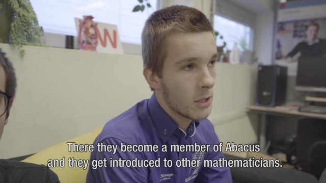
Wondering what it's like to study Mathematics? Kennet gives you a glimpse into his student life.
Why study Applied Mathematics?
- Use mathematics to solve practical and societal challenges
During this study programme, you will of course cover all the mathematical theory and skills you would expect from a bachelor’s degree in math. However, it does not end with that! This Bachelor’s has a strong focus on practical application, meaning that you will learn how to use your mathematical knowledge and skills to reduce complex, real-life issues to their mathematical essence, analyse them, and identify the best possible solution. You will discover that mathematics, like oxygen, is everywhere – and that you, as a mathematician, can offer solutions in many different societal domains, from the environment, mobility and technology, to insurance or healthcare.
- Cross disciplinary and geographic boundaries
At the University of Twente, we know complex societal challenges cannot be solved within one discipline. That is why as a student of Applied Mathematics, you will be encouraged to work not only with fellow mathematicians and engineers, but sometimes also with psychologists, communication professionals, public administration experts, philosophers or other social scientists. We believe interdisciplinary teamwork will help you discover the most relevant, ground-breaking solutions; besides, it will also prepare you for the work field after graduation. Furthermore, you will study with many foreign students and work with staff members from all over the world. So, if you are eager to cross both disciplinary and geographic boundaries in your future career, this Bachelor’s is the ideal preparation.
- Be highly confident in landing a good job
In more and more sectors, the demand for mathematicians is growing. The competencies you develop during your bachelor's in applying mathematics to practical challenges will make you highly sought after on the job market, especially when you have followed up your bachelor's with a master's. Do you want to challenge yourself even more, and broaden your chances on the job market? There is also the possibility of combining your study of Applied Mathematics with the Bachelor's in Technical Computer Science or Applied Physics (Dutch). Whatever path you choose, however, you can be confident in landing an interesting job soon after graduation!
- Attend a programme with a lot of personal attention
Education at the University of Twente is known to be personal and informal, and this Bachelor's is no different. We have an open-door policy and you will receive more individual attention and guidance than at most other universities. The atmosphere between lecturers and students is friendly and informal: you can always approach someone with your questions. Together with our active study association Abacus, we continuously improve our education and organise all kinds of study-related activities. Here, you will live your student life to the fullest and grow both professionally and personally!
Download brochure
Related stories
Future jobs
As a student of Applied Mathematics, you will have plenty of interesting job opportunities ahead of you. To give you an idea, we list some of the positions you could end up in as a graduate below.
- Data Scientist
- Operations Researcher
- Teacher
- Policy Maker
- AI Expert
- Research & Development Specialist
Programme overview
Certification: BSc
7522 NB Enschede
The Netherlands
Related programmes
Below you find a list of educational programmes which are (closely) related to Applied Mathematics.
Interested in science and technology, but rather not focus on one field? This Bachelor’s combines electrical engineering, mathematics, mechanical engineering, nanotechnology, robotics and more.
Learn to understand the fundamental laws of nature and apply them in innovative technologies and sustainable solutions in this Dutch-taught Bachelor’s.
Learn to design, develop and improve innovative healthcare technologies such as rehabilitation robots, artificial organs, prostheses and imaging technologies in this Dutch-taught Bachelor’s.
Use chemistry to understand, design and develop new sustainable processes, materials and solutions.
Roads, buildings, dikes, bridges: in this Bachelor’s, you learn how to combine technical and non-technical knowledge to design, construct and manage large projects in construction & infrastructure.
Want to use your creative ideas and AI for social impact? This Bachelor’s, in Enschede or Amsterdam, teaches you to create smart, responsible and innovative tech solutions to real-world problems.
In this unique programme, you will study Creative Technology in Amsterdam, while benefiting from the small scale, personal attention and technical expertise for which the UT is renowned.
Electrical engineering lies at the foundation of most technology. In this Bachelor’s, you dive into digital computing, wireless communication, and electronic sensors and systems to design the future.
Combine your creativity with technology to design and improve products, services and systems that make a real difference.
Are you analytical and interested in maths and business processes? In this Bachelor’s, you combine technical know-how, engineering, and management skills to improve business processes.
From rollercoasters to a robotic hand: in this Bachelor’s, available in Enschede and Amsterdam, you learn to invent, design and improve all kinds of appliances, machines, constructions and processes.
In this unique programme, you will study Mechanical Engineering in Amsterdam, while benefiting from the small scale, personal attention and technical expertise for which UT is renowned.
From programming and databases to networks and systems: use your fascination and talent for computers and technology to impact society and businesses.
Interested in science and technology, but rather not focus on one field? This Bachelor’s combines electrical engineering, mathematics, mechanical engineering, nanotechnology, robotics and more.
Learn to understand the fundamental laws of nature and apply them in innovative technologies and sustainable solutions in this Dutch-taught Bachelor’s.
Use chemistry to understand, design and develop new sustainable processes, materials and solutions.