Barnacle’s larvae leave behind tiny protein traces on a ship hull: but what is the type of protein and what is the protein-surface interaction? Conventional techniques can only identify dissolved proteins, and in large quantities. Using a modified type of an Atomic Force Microscope, scientists of the University of Twente in The Netherlands and A*STAR in Singapore, can now measure protein characteristics of even very small traces on a surface. They present the new technique in Nature Nanotechnology.
In infection diseases, membrane fouling, interaction with bacteria, as well as in rapid healing of wounds for example, the way proteins interact with a surface plays an important role. On a surface, they function in a different way than in solution. On a ship hull, the larvae of the barnacle will leave tiny traces of protein to test if the surface is attractive for long-term attachment. If we get to know more about this interaction, it will be possible to develop surface conditions that are less attractive for the barnacle. Large amounts of barnacles on a ship will have a destructive effect on flow resistance and will lead to more fuel consumption. The new measuring method makes use of a modified Atomic Force Microscope: a tiny ball glued to the cantilever of the microscope will attract protein molecules.
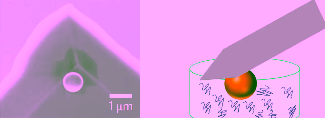
Modified AFM tip with a tiny ball that can attract protein molecules
FORCE MEASUREMENTS
An amount of just hundreds of protein molecules will be sufficient to determine a crucial value, called the iso-electric point (pI): this is the pH-value at which the protein has net zero electric charge. The pI value says a lot about the surroundings a protein will ‘feel comfortable’ in, and to which it preferably moves. Using the AFM microscope, of which the modified tip has collected protein molecules, it is possible to perform force measurements for different pH values. The tip will be attracted or repelled, or show no movement when the pI point is reached. For these measurement, the researchers made a special reference material consisting of several layers. Using this, the effect of a number of pH-values can be tested until the pI value is found.
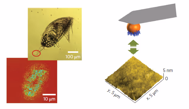
The traces the larve leaves behind (left) and force measurements (right)
PAINT CHANGE
The tests have been successfully performed for a number of known proteins like fibrinogen, myoglobine and bovine albumin. And returning to the barnacle: the tiny protein footprint will contain enough molecules to determine the pI value. This quantifies the ideal surface conditions, and using this knowledge, new choices can be made for e.g. the paint that is used on a ship hull.
The research has been done within the group Materials Science and Technology of Polymers of Professor Julius Vancso, in close collaboration with colleagues of A*STAR in Singapore – Prof Vancso is a Visiting Professor there as well. His group is part of UT’s MESA+ Institute for Nanotechnology.
The paper ‘Measuring protein isoelectric points by AFM-based force spectroscopy using trace amounts of sample’ by Shifeng Guo, Xiaojing Zhu, Dominik Janczewski, Serina Siew Chen Lee, Tao He, Serena Lay Ming Teo en Julius Vancso, recently appeared online in Nature Nanotechnology.
More recent news
 Thu 16 Jul 2026Veni grant for three UT researchers
 Wed 15 Jul 2026Globally unique measurements in Enschede to improve the prediction of urban heat stress
 Tue 14 Jul 2026UT professor Cristian Hesselman contributes to advice on strengthening the resilience of the Netherlands’ telecom and internet infrastructure
 Mon 13 Jul 2026From a Twente idea to a global company: Geert-Jan Bruinsma receives the Van den Kroonenberg Lifetime Achievement Award
 Thu 9 Jul 2026NWO grants awarded to four UT researchers