Last year, the University of Twente revealed the world’s most accurate 3D-printed biopsy robot. This year, during the Surgical Robot Challenge at the international Hamlyn Symposium in London, the researchers revealed the latest version. The Robot, named Sunram 5, is more accurate and faster, which means biopsy can be performed in an even better way.
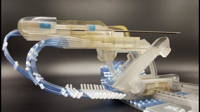
The Sunram 5 is driven by air-pressure-powered cylinders. Thanks to the use of hoses to supply the air, the controls of the robot can be placed outside of the MRI scanner. The design of the robot has been fully adapted to the current MRI setting. For instance, the robot is compact, which means it can be attached to the current breast compression system in various orientations. That way, the robot can easily reach any area in the breast. Another new feature are the so-called dual-speed motors, which allow for both faster and more accurate movements. This means accuracy has improved to 0.1mm, and the robot can move from start to its target location in about 10 seconds. Moreover, a safety mechanism has been designed which retracts the needle from the breast when the controller measures a mains voltage or air pressure failure.
Breast cancer
Breast cancer is the most common type of cancer among women. If a lesion, a piece of suspicious tissue, is found during the screening, further research is required to determine whether the tissue is benign or malignant. It is possible to retrieve a piece of tissue by placing a needle in the breast and navigating it towards the lesion. The tissue is sent to a pathologist, who can determine the nature of the tissue. The procedure described above is called a biopsy.
Accuracy
Accurate needle placement is essential for a biopsy. If the needle is in the wrong position, it is possible for a lesion to be found benign, while it is actually malignant. Not only biopsy procedures benefit from this accurate positioning, but cancer treatments that use the needle tip to destroy cancer cells benefit as well.
Robotics to the rescue
MRI scanners know no equal when it comes to locating lesions. Unfortunately, this quality is currently not fully utilised, as needle placement is performed manually. Robotics can play an important role here. However, not all robots can be used with MRI scanners. The scanner’s strong magnetic field means materials like metals cannot be used. That is why the UT, in cooperation with the ZGT (Ziekenhuis Groep Twente), developed a robot made entirely of plastic once before. The Sunram 5 is its successor.
UT scientists and partners
The development of Sunram 5 is in the hands of: Vincent Groenhuis MSc, Dr. Françoise J. Siepel, Marcel K. Welleweerd MSc, and Prof.dr.ir. Stefano Stramigioli of the Robotics and Mechatronics (RAM) lab at the University of Twente. Moreover, there was close cooperation with Dr. Jeroen Veltman, radiologist at the Ziekenhuis Groep Twente (ZGT), in order to adapt the design to clinical practice as much as possible. Abe van der Werf of Machnet B.V. was involved for the breast fixation system.
Image: Marcel K. Welleweerd MSc and Dr. Françoise J. Siepel


