In collaboration with various companies, scientists at MIRA research institute have recently developed a prototype for a handheld device that combines ultrasound technology with photoacoustics.
Doctoral candidate Pim van den Berg tested the technology for a variety of applications as part of his PhD research. His research shows that you can use the device to see a clear difference between arthritic fingers and healthy fingers. You can also use the technology to detect fibrosis of the liver in laboratory animals. In the future, fewer mice will probably be necessary in research into liver fibrosis. These first results are promising, but doctoral candidate Pim van den Berg believes additional research is necessary to help determine what conclusions can be drawn from what you see. However, the effect has been proven. The goal is to prepare the technology for applications throughout the world of medicine in the short term.
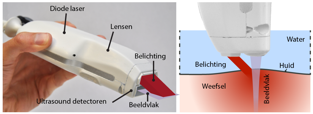
Where ultrasound offers images of structures, photoacoustics offers images that contain more functional information, such as where blood is located in the body. By combining both technologies in one device, you can generate images that offer considerably more information. The combination of ultrasound technology and photoacoustics was made possible through the integration of pulsating diode lasers in the ultrasound probe. The result is a compact, handheld and relatively inexpensive system.
Detecting arthritis
The combination of ultrasound and photoacoustics in a compact device means the applications of this technology include a simpler and more accurate diagnosis of the degree to which patients with rheumatoid arthritis are suffering from inflammation of the joints. Specialist Hein Moens from the ZGT is involved in the testing procedure: “I am very excited about this development and the opportunities this offers for the future. Current technologies only allow doctor’s to evaluate inflammation with the naked eye. This makes patients dependent on human perception. This technology will hopefully allow us to determine degrees of inflammation.
Researcher Pim van den Berg says “We have looked at fingers with and without inflammation using this device. The difference is clear. This method clearly shows the many extra blood vessels that form in the area of an inflammation. Additional research is needed to give us more information on these observations. We are now simply distinguishing extremes: inflammation, yes or no. The next step will be to identify the shades of grey in between.”
Laboratory animals with fibrosis of the liver
Fibrosis of the liver means damage to liver tissue, for example due to Hepatitis A or B, or alcohol. To date, no treatment is available. Scientists are using laboratory animals in research experiments to find potential new drugs to treat fibrosis of the liver. The device can help detect the disease in laboratory animals. This technology would allow researchers to track a single mouse for a longer period of time, for the purposes of discovering how the illness is progressing and learn more about the effects of medication. Fewer mice will be needed with the use of this technology. Professor Van den Berg comments that “We are now able to distinguish between fibrotic livers and healthy livers and that’s great, but further research is needed to help us determine what we can learn from what the technology is able to show us.”
Measuring blood velocity
The third application of this technology that Mr Van den Berg tested, is measuring the flow rate of blood. He worked with University College London on this part of his research. The flow rate of blood gives us more information about inflammation. Professor Van den Berg comments that “The test has been very successful. We would like to find out how fast the blood flows, how many blood vessels there are near the site of the inflammation and the levels of oxygen and nutrients. This information will tell us more about the inflammation. Our method allows you to measure the various components and the relationship between them. We were able to take excellent measurements in our laboratory environment. The next step is reviewing whether the device is able to do the same measurements on the human body.”
Effect
In photoacoustics, short laser pulses are emitted into a patient’s body. When the laser hits a blood vessel, for example, the light is converted into heat causing a small increase in pressure. This increase in pressure then moves through the body in the same way as a sound wave would. You can measure this soundwave on the skin. Photoacoustics technology is an extension of ultrasound imaging. In ultrasound imaging, the sound is transmitted into the body, where it bounces off of various tissues in a variety of ways and produces waves that can also be detected on the skin. Photoacoustics does not measure echo, but sound that is produced through the absorption of light. This means you need a much greater sensitivity to substances that absorb light, such as blood. The method is primarily suitable for measuring waves in relatively superficial parts of the body, up to 15 millimetres under the skin.
Future
In the future, the device could also be tested for use in mapping other ailments, such as skin cancer, burns or hardening of the arteries. “In a new European project with the same partners, we will be conducting measurements to greater depths to detect hardening of the carotid artery”, says Prof. Wiendelt Steenbergen, Pim van den Berg’s thesis supervisor.
Research
Pim van den Berg’s doctoral defence took place on 18 January 2017. His supervising professor is Dr Wiendelt Steenbergen. This research was conducted by the department of Biomedical Photonic Imaging at University of Twente’s MIRA institute in collaboration with Ruchi Bansal and Jai Prakash from the department of Biomaterials Science and Technology (MIRA) and with Thore Bucking, Joanna Brunker and Paul Beard from University College London. The study was partly funded by the European Commission (FP7/318067).


