Around a glioblastoma, a very aggressive brain tumor, cells of the human immune system start helping the tumor instead of attacking it. To do research on what happens in the interaction of these cells, scientists of the University of Twente now created a 3D-bioprinted mini model of the brain. Compared to existing lab models, the results using this 3D model match patient data much better. It will thus be a valuable way of testing new drugs, and it will reduce the number of animal trials. The research is published in ‘Advanced Materials’.
Immunotherapy, stimulating our own immune system in the treatment of cancer or other diseases, gets a lot of attention these days, and led to a Nobel Prize in 2018. At the same time, some immune cells show contrary action, they develop as accomplices of tumor cells. That is the case with the cells around a glioblastoma: so called macrophages, which infiltrate the tumor and help it spreading. Using the newly developed 3D model, a few millimeters in diameter, this mechanism can be studied far better than using 2D slices or using animal research: the results show a striking resemblance with actual patient data.
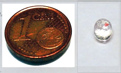
The size of the model, the red part is the tumor
Add and remove tumor
Thanks to developments in 3D bioprinting, the UT researchers could create a miniature brain model representing the delicate tissue around the tumor, including the macrophages. While printing, space was reserved for the tumor cells. It is possible to remove the tumor as a whole, for studying the effect on the remaining cells. The current model does not include nerve cells or blood veins: this would complicate the study of cell interaction a lot. It is, however, possible to add new cell types step by step, getting nearer to a realistic brain. In that case, one of the characteristics of glioblastoma, spreading like an octopus and thus getting very difficult to be removed operatively, may be demonstrated in a model as well.
Less animal trials
The basic ‘tumor micro environment’ (TME) that was printed now, already is a very valuable source of information. “For certain tumor markers, we see that they are up to a 1,000 times higher than we observe in 2D studies. This approaches realistic values far better”, says Marcel Heinrich. This clearly has an effect on medication as well: in the past, treatment that worked well in animals and 2D-lab experiments, sometimes failed in clinical trials. The 3D model shows why it isn’t working, because the dosage should be much higher, for example. One of the most challenging aspects is finding out if we can give the macrophages their original functionality, so they start attacking tumor cells again.
The promising results of a basic tumor microenvironment indicate that it can be used for other types of tumors as well. A major advantage is that this new 3D model will reduce the need for animal testing.
Marcel Heinrich is a PhD student within the section Targeted Therapeutics of Prof. Jai Prakash, part of the Biomaterials Science and Technology group, TechMed Centre.
This research fits well within the UT theme ‘Improving healthcare by personalized technologies’
The paper ‘3D-Bioprinted Mini-Brain: A Glioblastoma Model to Study Cellular Interactions and Therapeutics’, by Marcel Heinrich, Ruchi Bansal, Twan Lammers, Yu Shrike Zang, Raymond Michel Schiffelers and Jai Prakash, appears in Advanced Materials


