what we do
We investigate the use of light for medical purposes. Our aim is to develop optical and hybrid optical-acoustical technologies for medical research and diagnosis, in particular in the fields of oncology, wound healing and microscopy. Physiological properties of primary interest to us are microcirculatory blood flow, hemoglobin concentrations, blood oxygenation and scattering properties in general.
Our approaches include physical research into light-tissue interaction and its measurement, biomedical engineering to realize suitable instrumentation for ex/in vivo use, and clinical evaluation together with several medical partners.
Our research topics
- Wavefront shaping
Scattering of light forms a huge limitations for optical imaging, but also severely limits telecommunication, spectroscopy, and other optical techniques. In the last few decades, a tremendous effort was put in developing imaging methods that work inside turbid biological tissue. That research has brought forward important new imaging methods like optical coherence tomography, diffusion tomography and laser speckle velocimetry.

- Flow measurement with dynamic optical speckles
If you see laser light reflected from a rough surface or scattering object, you observe tiny dots on your retina: the so-called speckles. If particle move within the scattering object, such as red blood cells in tissue, the speckles move too. This phenomenon is the basis of Laser Doppler Perfusion Imaging (LDPI) and Laser Speckle Contrast Analysis.
- Human milk and lactation
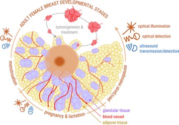
Breastfeeding has unique and unparalleled advantages for mothers and infants: it supports optimal physical and cognitive development, it protects against infections, and it plays an important role in the development of our microbiome[i]. But despite all of these benefits, many mothers experience breastfeeding problems, and many aspects of human milk and lactation are not yet completely understood[ii].
We use our optical technologies to learn more about the composition of human milk and the physiology of lactation. The majority of our research is currently funded by a Starting Grant from the European Research Council (ERC): LactIns-and-outs.
- Optical coherence tomography
Premature and ill newborns are often subjected to invasive blood sampling. Invasive blood sampling is related to pain, stress, substantial blood loss and increased risk of infections – all factors that have been proven to impair the growth and development of this vulnerable patient group.
A possible alternative to invasive blood testing is optical spectroscopy: a fast, noninvasive technique, sensitive to the concentration dependent light absorption by two frequently – often exclusively – tested blood analytes in newborns (bilirubin and hemoglobin). Transcutaneous bilirubin meters (TcB meters) are devices based on optical spectroscopy, which are currently applied in the clinic to noninvasively measure bilirubin levels in the skin of newborns. This method is currently only used for screening purposes. TcB meters can prevent around 30% of all invasive blood samples for bilirubin determinations. For the other 70%, the technique is unfortunately not accurate enough to replace invasive blood sampling. The main reason is that the skin concentration of bilirubin is not directly related to the blood concentration (TSB), which is used for medical decision making.
We investigate how we can improve the clinical value of TcB meters - in close collaboration with the Isala Hospital. In parallel, we investigate how we can use another optical technique - optical coherence tomography - to potentially perform a more accurate noninvasive blood analysis.

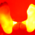
Image: OCT B-scans of muscular tissue (left) and fatty tissue (right).
- Photoacoustic imaging
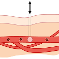
Photoacoustic (optoacoustic) imaging is one of the most exciting methods under investigation for imaging soft-tissues. The method uses light pulses as the probing energy beam, with the aim to visualize sites where optical absorption takes place in tissue. However, photons are not detected as the signal of interest, as is typically done in x-ray imaging. This is because light scatters considerably in tissue, and the sharp shadows seen in x-ray imaging will not be observed using light.
The localization of absorption is done, with the knowledge that light absorbed is converted into heat, and the resulting temperature rise causes thermal expansion. When short pulses of light are used, the thermal expansion results in a mechanical wave being generated originating at the locations where optical absorption took place. The mechanical wave has frequency components in the ultrasound regime, and can be detected at the boundary of the tissue using ultrasound detectors. From this point on the ultrasound source positions can be located using methods well known in ultrasound imaging.
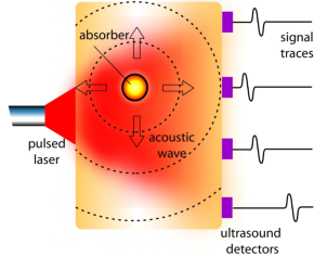
To summarize: The mechanism of pulsed photoacoustic signal generation consists of the following steps:
- Thermography
Thermography is the imaging of the temperature of the surface of objects. Physiological changes in the human body are often accompanied with a change in temperature. Therefore a thermal sensor or camera be used as a monitoring device for certain processes such as inflammation.
You want to help BMPI in their research?
- Projects, Master and Bachelor
We are always on the lookout for enthusiastic students to contribute to our ongoing main projects. Whether you are a bachelor or master student in Biomedical Optics, Technology or Applied Physics, we have exciting projects that will provide you with hands-on experience in cutting-edge research. If you have a different background and you think you can be a great asset for one of our projects, let us know!
Join our team and work alongside experienced researchers to explore the latest advancements in biomedical optics, and use this knowledge to develop innovative solutions for medical diagnostics.
- Vacancies
Are you looking for an opportunity to further your research career? We are seeking talented and driven individuals to join our team and help drive our ongoing projects forward. Check out our vacancies page for possible vacancies in our group.
