Introduction to Photoacoustic Imaging
Bring me to the available projects!
Photoacoustic (optoacoustic) imaging is one of the most exciting methods under investigation for imaging soft-tissues. The method uses light pulses as the probing energy beam, with the aim to visualize sites where optical absorption takes place in tissue. However, photons are not detected as the signal of interest, as is typically done in x-ray imaging. This is because light scatters considerably in tissue, and the sharp shadows seen in x-ray imaging will not be observed using light.
The localization of absorption is done, with the knowledge that light absorbed is converted into heat, and the resulting temperature rise causes thermal expansion. When short pulses of light are used, the thermal expansion results in a mechanical wave being generated originating at the locations where optical absorption took place. The mechanical wave has frequency components in the ultrasound regime, and can be detected at the boundary of the tissue using ultrasound detectors. From this point on the ultrasound source positions can be located using methods well known in ultrasound imaging.
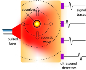
To summarize: The mechanism of pulsed photoacoustic signal generation consists of the following steps:
- Light is selectively absorbed in higher absorbing regions, when the investigated volume is exposed to pulsed laser radiation;
- Fast non-radiative relaxation of excited states takes place with thermalization of absorbed optical energy;
- The resulting local thermal expansion produces pressure transients.
The pressure pulses propagate as ultrasound to the surface where it is detected using wide-frequency band detectors. The time-of-flight, amplitude and duration of the pressure transient generated under irradiation conditions of pressure and thermal confinement provide information regarding location, strength and dimensions of the acoustic source, thereby permitting a reconstruction of the absorber.
The method thus combines the advantage of using light which shows high absorption contrast by hemoglobin in blood, with the advantage of detecting ultrasound namely low scattering and attenuation in tissue. Further advantages in the use of light are that the red and near-infrared wavelengths used constitute non-ionizing radiation, and also provide the capability to do spectroscopy and possibly identify more physiological information from tissue.
Typically photoacoustics is used is visualizing the blood vessels which are abnormal in form and amount in pathologies such as cancer and inflammation, or in imaging other absorbers for example melanin which is prominent in melanomas.
Research orientations in the group
In the group we have pioneered work in photoacoustic imaging of the breast using a research imager (Figure 1) approved for use in women. This work which we call PAMmography (Photoacoustic Mammography) is being done in collaboration with radiologists at the Medisch Spectrum Twente (Oldenzaal).
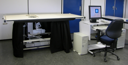
Figure 1 The Twente Photoacoustic Mammoscope.
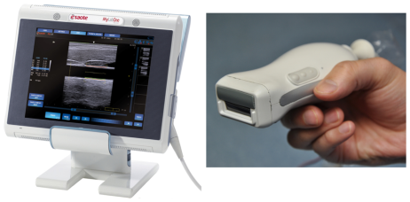
Figure 2 Portable imaging scanner combining photoacoustics and ultrasound, left is the ultrasound scanner system, right is the picture of the probe integrating laser module and ultrasound transducer array.
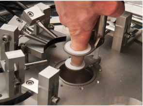
Figure 3 Photograph of the photoacoustic computed tomography (PA-CT) setup. The finger is maintained stationary in water at the center of rotation of the curvilinear array, while illumination is provided by optical fibers in a backward-mode configuration.
Further, we apply two variants (Figure 2 and 3) of photoacoustic imagers to visualize indicators of inflammation associated with rheumatoid arthritis in collaboration with rheumatologists at the Ziekenhuisgroep Twente (Almelo). Further we also work in studying various optical and physical properties of biological materials in laboratory conditions using the photoacoustic method.
Contact:
- Srirang Manohar (s.manohar@utwente.nl) for information regarding PAMmography (Photoacoustic Mammography)and the use of Photoacoustic Computed Tomography for finger joint imaging;
- Wiendelt Steenbergen (w.steenbergen@utwente.nl) for information regarding the use of Photoacoustic-Ultrasound linear mode imaging in finger joint imaging, and for optical property and Gruneisen property measurements using photoacoustics
Selected References:
[1]Heijblom, M. H., Steenbergen, W., & Manohar, S. (2015) Clinical Photoacoustic Breast Imaging: The Twente Experience IEEE Pulse, in print. (May-June issue)
[2]Daoudi, K., van den Berg, P. J., Rabot, O., Kohl, A., Tisserand, S., Brands, P., & Steenbergen, W. (2014). Handheld probe integrating laser diode and ultrasound transducer array for ultrasound/photoacoustic dual modality imaging. Optics express, 22(21), 26365-26374.
[3]Villanueva, Y., Hondebrink, E., Petersen, W., & Steenbergen, W. (2014). Photoacoustic measurement of the Grüneisen parameter using an integrating sphere. Review of Scientific Instruments, 85(7), 074904.
[4]Langhout, G. C., Grootendorst, D. J., Nieweg, O. E., Wouters, M. W. J. M., Hage, J. A. V. D., Jose, J., Van Boven, H., Steenbergen, W., Manohar, S. & Ruers, T. J. M. (2014). Detection of melanoma metastases in resected human lymph nodes by noninvasive multispectral photoacoustic imaging. Journal of Biomedical Imaging, 2014, 5.
[5]van Es, P., Biswas, S. K., Moens, H. J. B., Steenbergen, W., & Manohar, S. (2014). Initial results of finger imaging using photoacoustic computed tomography. Journal of biomedical optics, 60501, 1.
[6]Daoudi, K., Van Es, P., Manohar, S., & Steenbergen, W. (2013). Two-dimensional spatiotemporal monitoring of temperature in photothermal therapy using hybrid photoacoustic-ultrasound transmission tomography. Journal of biomedical optics, 18(11), 116009-116009.
[7]Xia, W., Piras, D., Van Hespen, J., Van Veldhoven, S., Prins, C., Van Leeuwen, T., van Leeuwen, T. G., Steenbergen, W., & Manohar, S. (2013). An optimized ultrasound detector for photoacoustic breast tomography. Medical Physics 40, 032901.
[8]Piras, D., Grijsen, C., Schütte, P., Steenbergen, W., & Manohar, S. (2013). Photoacoustic needle: minimally invasive guidance to biopsy. Journal of biomedical optics, 18(7), 070502-070502.
[9]Heijblom, M., Piras, D., Xia, W., van Hespen, J. C. G., Klaase, J. M., van den Engh, F. M., van Leeuwen, T. G., Steenbergen, W., & Manohar, S. (2012) Visualizing breast cancer using the Twente Photoacoustic Mammoscope: What do we learn from twelve new patient measurements? Optics Express 20, 11582.
[10]Piras, D., Xia, W., Steenbergen, W., van Leeuwen, T .G. and Manohar, S. (2010) Photoacoustic Imaging of the Breast Using the Twente Photoacoustic Mammoscope: Present Status and Future Perspectives. IEEE Journal Selected Topics Quantum Electronics 16, 730.