Light tissue interactions and their measurement
Our technologies make use of the absorption of light within the tissue, and the Doppler shift when light is scattered by moving objects.
We concentrate the following technologies:
Photoacoustic imaging
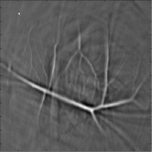
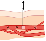
Photoacoustic imaging is ultrasound imaging, where the ultrasound is created by short light pulses. These short pulses are absorbed by blood in tissue, which creates ultrasound. Based on this ultrasound, the presence of blood can be imaged in 3D. We develop this technology for small animal imaging, imaging superficial tissues like skin, and the detection of breast cancer.
For more information click here.
Acousto-optic imaging
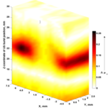
In acousto-optic imaging, tissue is irradiated with light and focused ultrasound at the same time. Light that travels through the ultrasound focus is ‘labeled’ by the acoustic vibrations, and this labeled light can be detected outside the tissue. In this manner, the distribution of light within tissue can be measured.
For more information click here.
Wavefront shaping
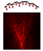
Using a technique called wavefront shaping, it is possible to sharply focus light deep inside biological tissue. We are developing new methods to use wavefront shaping for high-resolution imaging inside non-transparent tissue.
For more information click here.
Flow measurement with dynamic optical speckles
 If you see laser light reflected from a rough surface or scattering object, you observe tiny dots on your retina: the so-called speckles. If particle move within the scattering object, such as red blood cells in tissue, the speckles move too. This phenomenon is the basis of Laser Doppler Perfusion Imaging (LDPI) and Laser Speckle Contrast Analysis.
Contrast enhancement of photoacoustics using gold nanoparticles
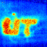
We developed gold nanorods (GNR) which strongly absorb near infrared light. We make the nanorods sensitive to tumor cells by binding antibodies to the rods. We investigate the physical processes when the nanorods are heated by short light pulses, and the use of GNRs as photaocoustic contrast agent and as tool for therapy.
Spectroscopic Optical coherence tomography
 Optical Coherence Tomography (OCT) is a high resolution, noninvasive 3D-imaging technique employing light. Spectroscopic OCT (sOCT) is one of the functional extensions of OCT, which can be used for depth-resolved spectroscopy in tissue. At BMPI, we develop sOCT specifically for the noninvasive analysis of blood composition in newborns.