For the first time in millennia, we can analyse why a vaginal pessary remains in place. UT scientist Frieda van den Noort (TechMed Centre / group M3I) and her colleagues recently published this insight in Nature Scientific Reports. With this examination, a huge number of women, suffering from pelvic organ prolapse, can be helped who are now dependent on surgery or who 'have' to learn to live with their complaints. For example, a standing MRI scan provides insights and enables personalised pessaries in 3D prints.
"With the results of this study, we can analyse, thanks to a standing MRI scan, why a vaginal pessary remains in place. This provides opportunities to redesign the pessary. With the current possibilities in 3D printing, we can work towards personalised pessaries, so that we can increase the success of pessaries. With this, we can help a huge number of women who are now dependent on surgery or 'have' to learn to live with their complaints."
Pelvic organ prolapse
"The most commonly used pessary is a ring. But until now, it was unknown why the pessary remains in the vagina. This question is relevant because pelvic organ prolapse is a common problem (40% of all women over the age of 40). Pessaries are the only treatment options besides pelvic physiotherapy that can prevent the need for surgery. Unfortunately, the success rate of pessary measurements is only about 63%. To increase this success, we need to know which mechanisms influence a pessary measurement. We can then adjust the design of the pessary and increase the success rate."
Upright scanning in MRI
“Until now, it was not possible to properly examine pessary measurements, because with MRI imaging, the patient is scanned lying down. This is even though pessary measurements often fail, because the patient loses the pessary in a standing or sitting position, in other words under the influence of gravity. At the UT, however, we have an MRI scanner in which we can scan the patient in an upright position. We used this scanner to scan thirty patients with a successful pessary measurement and see how it is supported when the patient is standing."
Statements
"Over time, several hypotheses have been formulated in the literature about why a pessary remains in place, which we have confirmed or disproved with the help of the MRI images."
1. The main hypothesis was that the pessary remains behind the pelvic floor bone (symphysis). We were able to disprove this hypothesis because the bottom of the pessary is below the symphysis in all women.
2. The second hypothesis was that the pelvic floor muscles support the pessary. By analysing the position of the muscles to the pessary on the MRI images, we were able to conclude that this hypothesis is true. In all cases, the pessary rests neatly in the pelvic floor muscles 'hammock'.
3. The last hypothesis was that the uterus holds the pessary in place, this hypothesis is probably also true, because in almost all cases the cervix is in the middle of the pessary. As a result, the uterus presses the pessary against the vaginal posterior wall, preventing it from slipping out easily.
Follow-up research
Follow-up research has now also been done on thirty women with an unsuccessful pessary measurement and the results are available for peer review in a scientific journal. This research confirms the findings in the current work.
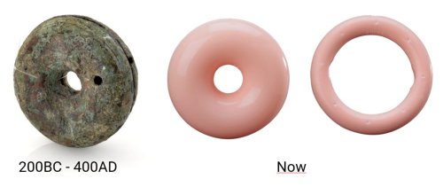
Photo: © The Board of Trustees of the Science Museum.
History of vaginal pessaries
The Egyptians and Romans already used vaginal pessaries against pelvic organ prolapse in women. The Egyptians used pomegranate peels and a bronze pessary has been found from Roman times that shows great similarities with the donut pessary that is still used today (see image).
Technology for Women’s Health
Aiming to optimize and personalize women’s health across the entire life cycle, a multidisciplinary team of researchers at the University of Twente has formed a new cluster: Technology for Women’s Health. The group conducts research on women’s physical and mental health, their specific healthcare needs, and overall well-being. This research aims to understand and explain why differences between men and women arise in the manifestation of health and disease, as well as why women often respond differently to treatments. At its core, the focus is on developing or adapting technologies that can be applied to prevention, diagnosis, treatment, and aftercare.
The article in Nature Scientific Reports can be found here.


