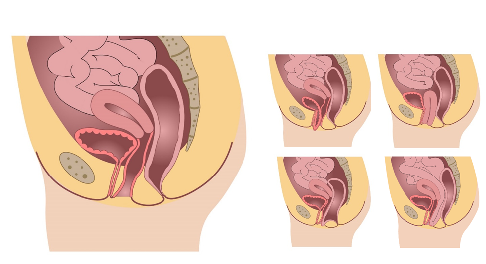
Many women, mainly postmenopausal and with a history of vaginal delivery, suffer from pelvic organ prolapse (POP). POP is described as a downward displacement of one or more of the pelvic organs, including the bladder, uterus, bowel and rectum (Figure below). As a consequence, symptoms such as vaginal bulging, pelvic pressure and incomplete emptying of the bladder or defecation difficulty may occur. Therapeutic options are found in pessary (ring) placement, pelvic physiotherapy and different types of surgery.
The diagnosis of the extent of prolapse is generally estimated with the patient in supine (lying down) position, while women experience the most complaints in standing position. Research using the low-field MR tilting system or transperineal ultrasound gives several new possibilities:
1. Providing insight in the extent of prolapse in different patient positions;
2. Estimate the effect of pessary and surgical treatment;
3. Improve and tailor-make diagnosis and therapy.
4. Diagnose musculature pathology (e.g. muscle avulsion)
The links below provide background on several ongoing research projects:
EPPA: Understanding the effect of a pessary on the pelvic anatomy (upright MRI, segmentation)
TORBO: Studying recurrences after prolapse surgery (upright MRI and ultrasound)
FIT-UP: Towards tailor-made pessaries (MRI, ultrasound, EMG, FEM, sensors)
GYNIUS: Functional ultrasound assessment of the pelvic floor muscles (US, segmentation, strain)
If you are interested in a master or bachelor assignment related to the above mentioned topics, please contact Anique Bellos-Grob (a.t.m.bellos-grob@utwente.nl).