In order to detect an influenza virus successfully, even in small concentrations, you would like to know in what way the virus interacts with healthy cells. Researcher Mark Verheijden of the University of Twente succeeded in mimicking the cell surface on a sensor. In this way the virus is detected in a dynamic environment. This bio-inspired layer can also be used as a biocompatible coating for implants, for example.
To detect the presence of a virus is one thing, but this doesn’t give us much information about the way it affects healthy cells. Whenever we use tests for virus prevention, we need to know how this interaction works. In this way it is also possible to see differences in interaction for humans and animals. The classic detection method is not based on this interaction and the dynamics that go with it. Instead, capturing a virus on a surface that behaves like a cell membrane is preferable. Verheijden succeeded in mimicking this surface.
Bird flu
The layer is a bilayer of lipids: soap molecules that can behave like butter or oil. Verheijden used this basic bilayer to study the effect of Influenza A. This virus can have different effects on humans or birds, for example. And there the interaction with healthy cells comes in: once the virus mutates, it may get dangerous for humans as well. The virus detects specific sugar structures at the cell surfaces and attaches to it. Verheijden added these sugars to the bilayer. As a model for the virus, he chooses hemagglutine (HA), this is the surface protein that binds the virus. This is a new approach; much is known on viruses, but the actual interaction with cells and the dynamics of their membrane is still not well understood and an area of research.
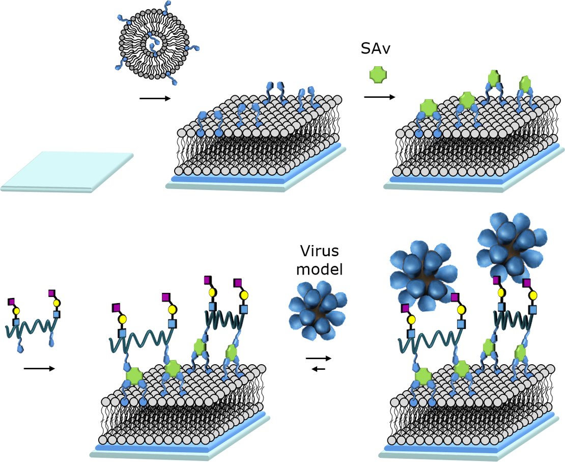
Lipid bilayer interacting with a virus model (in blue)
Organ-on-a-chip
The bioinspired layer can be used for other purposes as well. Implants, for example, can be made biocompatible and show less . In other cases, the layer can be used to keep the surface as clean as possible. On a catheter surface, you would like to interaction. The bilayer can also detected so-called vesicles, as biomarkers in disease processes. Furthermore, in the rapidly evolving field of organ-on-a-chip systems, the layers are applicable for creating real-life circumstances for the miniature organs.
The lipid bilayer structure is a powerful platform for research on biological interactions and the way these can be steered in a direction. The actual measurement is done by using optical techniques as well as high-precision weighing equipment like quartz crystal microbalance.
Verheijden did his research in the Bioinspired Molecular Engineering lab, led by Prof Pascal Jonkheijm. He did his work in close cooperation with researcher Daniele di Iorio. The lab is part of the MESA+ Institute for Nanotechnology and the TechMed Institute, both of the University of Twente. The work was made possible through the Vidi-grant that Pascal Jonkheijm received from the Netherlands Organization for Scientific Research (NWO).
Mark Lloyd Verheijden (Oosterhout, 1989) defended his PhD thesis ‘Supramolecular binding of vesicles, viruses and cells to biomimetic lipid bilayers’ on 25 May 2018.


