Welcome to the BioImaging Centre
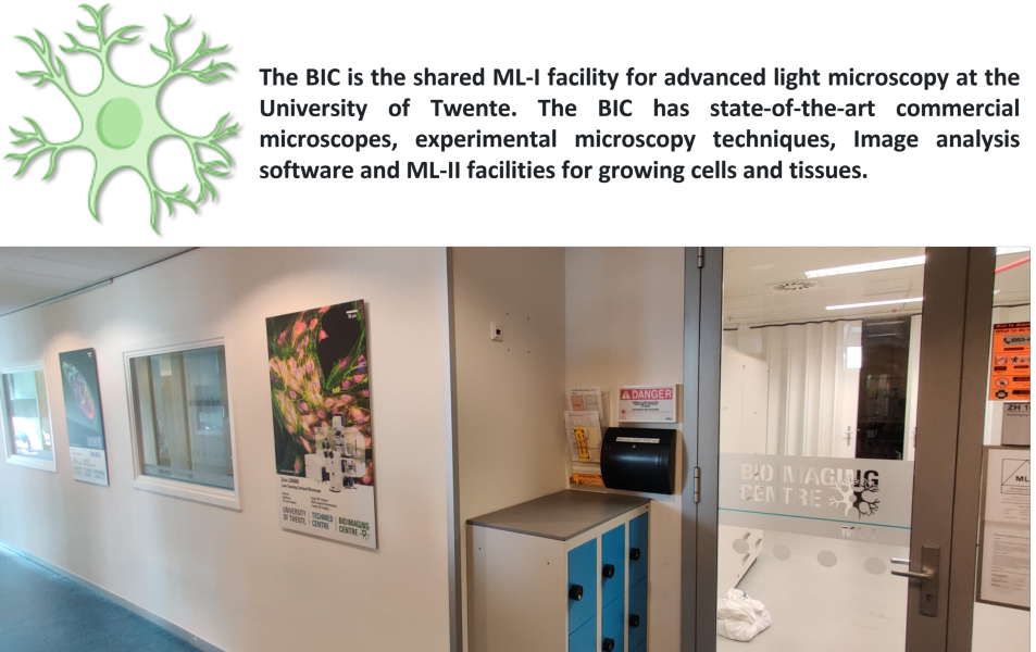
What the BIC has to offer
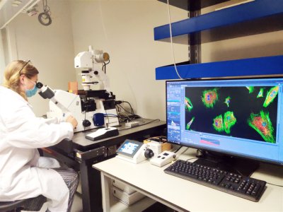
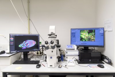
- State-of-the art microscopes
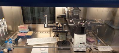
- Laser scanning confocal fluorescence
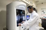
- Zeiss LSM880
The Zeiss LSM880 confocal microscope is a powerful imaging tool used in biological and materials science research. It utilizes laser scanning technology to generate high-resolution, three-dimensional images of samples, allowing for the visualization of subcellular structures and fine details of tissues and materials. The LSM880 confocal microscope can be used for a wide range of applications, including fluorescence imaging, live-cell imaging and spectral imaging.
- Opera spinning disk
The Opera spinning disk is an advanced imaging system that utilizes a spinning disk confocal microscope to capture high-resolution images of biological samples.
This system is capable of high-speed, multicolor imaging of live cells and tissues, allowing researchers to study dynamic processes such as cell division and migration.
- Brightfield / Widefield fluorescence
- Zeiss Axio Observer
The Zeiss Axio Observer is a versatile microscope that can perform a range of basic imaging techniques such as brightfield, darkfield, phase contrast, and widefield fluorescence microscopy. This system is ideal for users who want to quickly visualize their samples.
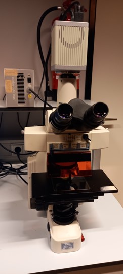
- Nicon Eclipse E400
The Nikon Eclipse E400 microscope is suitable for biological and materials science applications. It provides detailed imaging of samples and features advanced capabilities such as phase contrast and fluorescence microscopy for studying live cells and tissues.
The microscope has a camera connected to capture your images, very usefull together with our available Imaris image analysis software.
Overall, the Nikon Eclipse E400 is a versatile tool for scientific research for a variety of applications.
- Holotomography
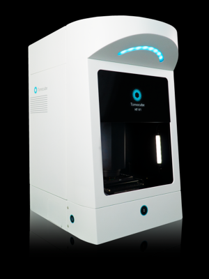
- Tomocube
Observations of cellular morphology and activity often rely on labeling target molecules which can be invasive, alter the target molecules, and lack quantitative information. Common imaging tools, such as laser-based microscopes, may also cause phototoxic damage to cells. Holotomography (HT) imaging can visualize cells without labeling by capturing the light scattering properties of cellular materials using low levels of light intensity. The Tomocube HT-X1 Holotomography can collect and selectively color refractive index information to reveal cells and their organelles, while also providing quantitative 3D data such as volume, surface area, and dry mass.
- Raman
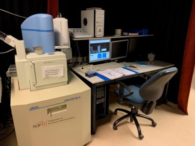
- SEM-Raman (785nm)
The SEM-Raman microscope is a powerful analytical tool that combines two distinct imaging techniques - scanning electron microscopy (SEM) and Raman spectroscopy (at 785nm)
SEM provides high-resolution imaging of sample surfaces, while Raman spectroscopy provides detailed chemical information about the sample composition.
This combination enables researchers to study the morphology and composition of materials with a high degree of accuracy, making it useful for applications such as materials science and nanotechnology research.
Furthermore, the SEM-Raman microscope can be used to study a wide range of materials, including biological samples, polymers, semiconductors, and more. Overall, it is a valuable tool for characterizing the properties of complex materials and investigating their structure-function relationships.
- Raman (640nm)
- Raman (405nm)
- Witec alpha 300 Raman (532, 633, 785nm)
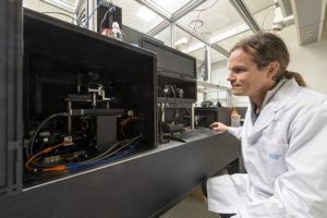
- Experimental microscopy
Next to commercial microscopes we also work on novel microscopy techniques to visualize even more!
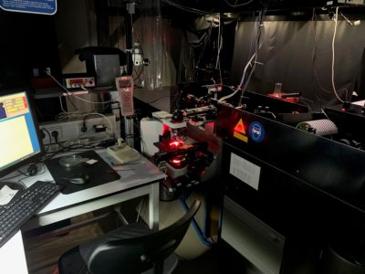
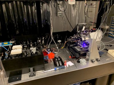
- Wavefront-shaping 2-photon microscope
Our homebuilt Two-Photon laser scanning microscope is a powerful imaging tool used in biological and neuroscience research. It uses high-intensity infrared laser pulses to excite fluorescent molecules in living tissues, allowing for the visualization of fine details deep within the sample. Compared to traditional confocal microscopes, it can image deeper and with less photodamage. This microscope is ideal for studying the structure and function of living tissues, including the brain, and organs on a chip systems.
Additionaly our Two-Photon laser scanning microscope includes a wavefront shaping module which can correct for tissue distortions and sample abberations which allows for deeper imaging of biological samples. This module enhances the microscope's capabilities, enabling researchers to visualize previously inaccessible structures and processes within living tissues.
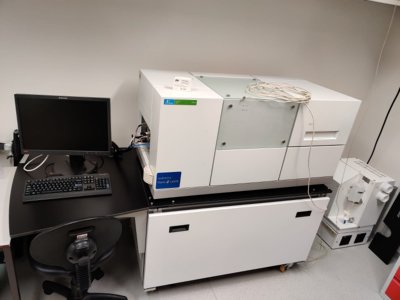
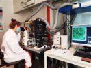
- Single cell analysis systems
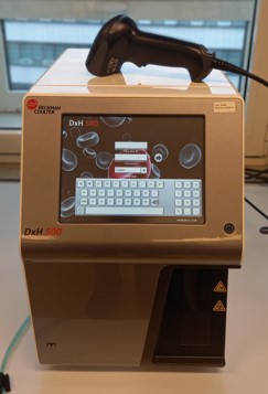
- Hematology Analyzer
The Beckman Coulter Hematology Analyzer is a medical device used to perform complete blood count (CBC) tests, which measure the number and characteristics of blood cells in a patient's sample.
This analyzer uses a combination of electrical impedance and laser-based technology to rapidly analyze blood samples.
The analyzer can measure a wide range of blood parameters, including red blood cell count, hemoglobin levels, white blood cell count, and platelet count.
- VyCAP cell puncher
The VyCAP Single-cell Analysis System is used to isolate and analyze individual cells from biological samples.
This system uses microfluidic technology to sort and manipulate single cells, allowing for high-throughput analysis of their genetic and phenotypic characteristics.
The VyCAP system is capable of performing a range of assays, including PCR, sequencing, and protein analysis, to generate comprehensive information about individual cells.
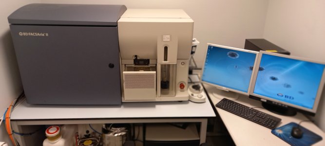
- FACSAria II Flow Cytometric Cell Sorter
The BD FACSAria II Flow Cytometer is a state of the art device that allows users to analyse and sort cells and particles in micrometer range (1-50um) based on their physical properties like size and internal structure as well as fluorescence labeling.
It features 3 lasers (375nm UV, 488nm blue and 633nm red) and 13 sensitive PMT (PhotoMultiplier Tube) detectors making mulitplex staining and analysis of samples possible.
Cells or particles of interest can be defined using gates and instantly be sorted in any number from a single to a million of cells into 5ml tubes, 6/12/24/96/384 well plates or regular microscope slides (76x26mm).
Analysis and sorting of cells/particles can be achieved with a very high throughput of up to 20000 cells per second, making it a great device for analysing suspended particles.
Data analysis can be done with dedicated commercial software (DIVA 7.0), but data can also be exported and analysed in other freeware software or Matlab using a flowcytometry Matlab script.
Calibration beads for quantifying the sample volume, size or intensity of particles are available upon request.
In summary our flowcytometer can be of great help during your student or PhD research in analysing and characterizing your (biological) samples.
Sample Requirements:
- Concentration should be about 1 million cells/particles per milliliter
- A minimum sample volume of 100ul is needed
- Always bring an unstained negative control
For further questions ask Christian Breukers: c.breukers@utwente.nl
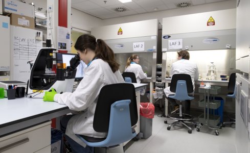
- Cell culture facilities
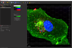
- Image analysis software
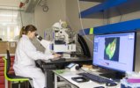
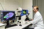
The BIC also offers image analysis software to analyze your data, free of charge. Our first available software package is Imaris.
This software can be used on the stand alone Imaris PC in the BIC lab (ZH160) or accesed remotely via "Remote Desktop".
If you want to make use of this software, please send an E-mail to:
You would like to use one of our systems, now what?
We always try to assist researchers in addressing complex imaging challenges. We provide training and support to users of our imaging systems, ensuring that users have the skills and knowledge needed to operate the equipment effectively.
In addition, our technicians are available to troubleshoot issues and provide guidance on experimental design. For researchers who require more advanced imaging solutions, our technicians can also work with them to develop customized imaging protocols and techniques.
The BIC is available for both the University of Twente and external users.
The BioImaging Centre is a TechMed facility and affiliated with the NL-BioImaging AM distributed infrastructure.