Nanobiophysics Group uses two main laboratories, the physics laboratory hosts many modern tools to study cell interaction and protein aggregations. Many (custom-build) microscopes are available build for a broad and versatile applications. The microscopy lab ZH174 is restricted area due to the AP-I & LASER classification 3b, 4. It's allowed to study ML-1 calssified samples in a closed container.
Location [Building 28] Zuidhorst: 1.74
Laboratory contact and safety manager is Robert Molenaar
Keywords: Nanopositioners, lasers, fluorescence, TIRFM total internal reflection microscopy, darkfield, AFM atomic force microscopy, confocal microscopy, fluorescent lifetime, spectroscopy, lightscatter. nanophotonics, LDOS, imaging,
Overview of microscopy techniques that are available.
Atomic Force microscopy.
- Bruker Catalyst AFM for scanning, tapping and peak force imaging, compatible with live cells.
- Multiple top-head design AFM systems, for versatile sample interaction/manipulation by AFM.
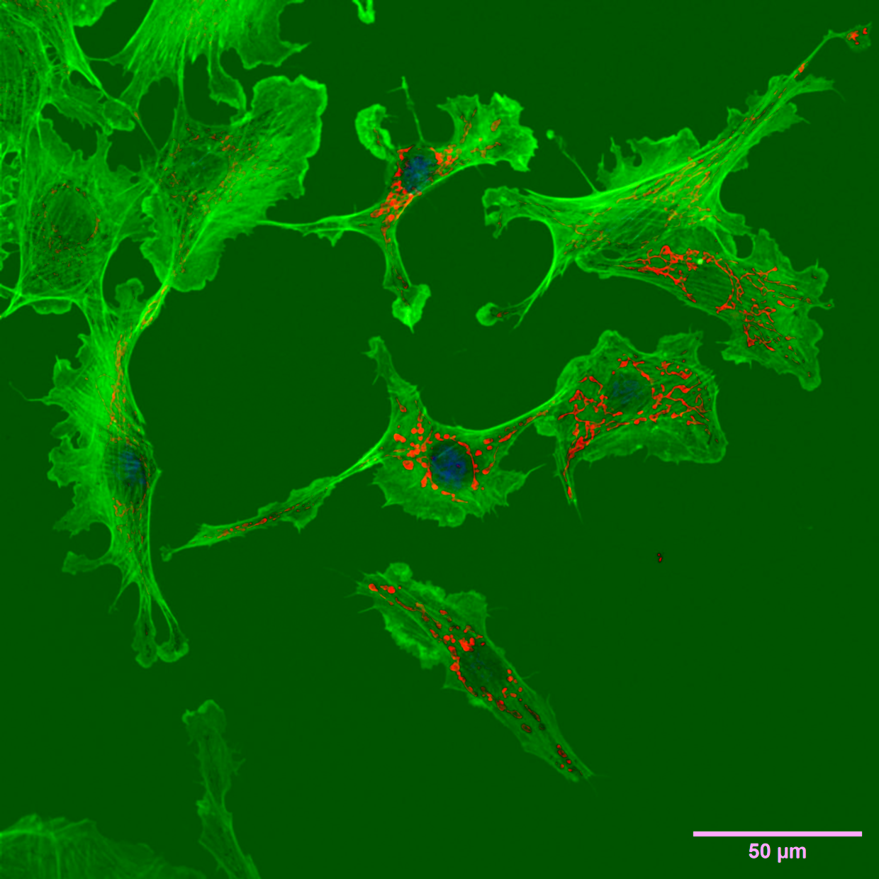
FluorescenT Microscopy Techniques
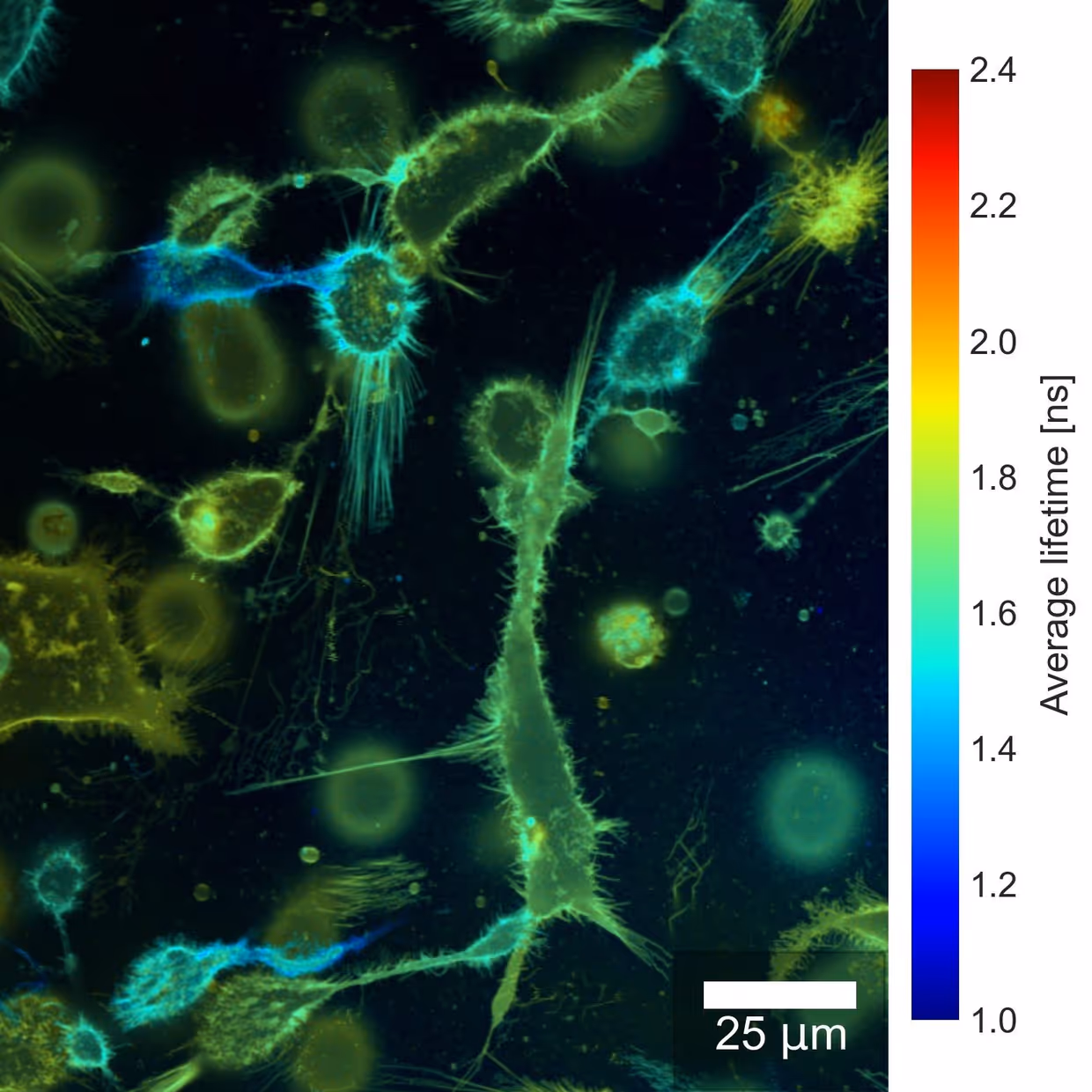
- PicoQuant MT200 Laser scanning confcoal microscope with 4 channels: single molecule sensitive applications, confocal imaging, FCS, FCCS, FLIM, fluorescent lifetime, phosphorescence Lifetime, FRET burst analysis, anistropy, antibunching and nanosecond FCS.
- PicoQuant, FCS compatible microscope, dual TCSPC channels.
- PicoQuant, FCS compatible microscope, dual TCSPC channels.
- Confocal microscope with TCSPC for active Local Density of States modification.
- Fianium pulsed light source, full white light spectrum.
- Nikon Ti-E TIRF microscope, wide field based for PALM, STORM dual channel widefield/TIRF.
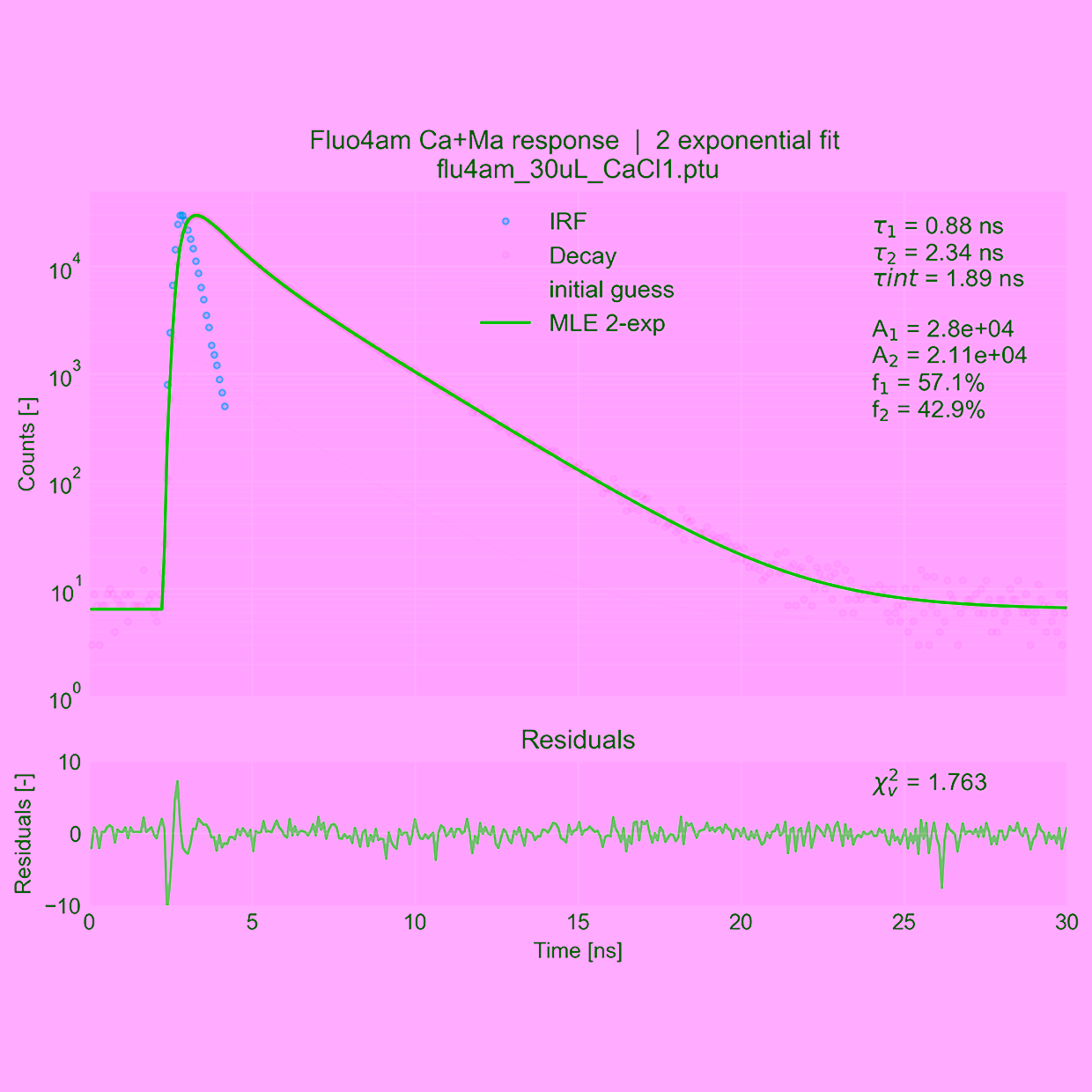
- Fluoromax-4 fluorospectrometer with lifetime extension.
- Zeiss Axioivert fluorescent microscope.
- Nikon TI-2000 fluorescent microscope with live cell incubator.
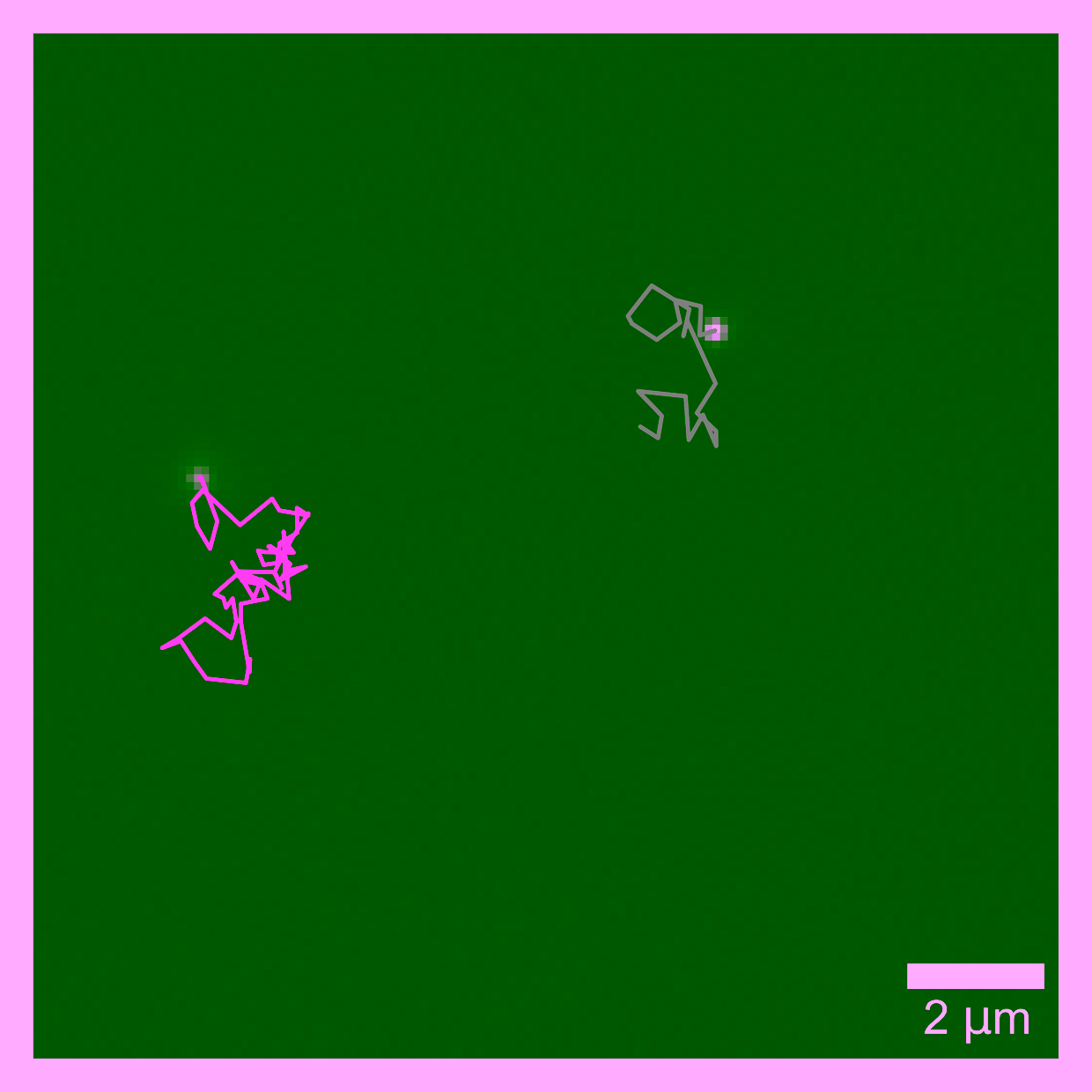
- Nikon TI-2000 Laser excited fluorescent microscope for single particle tracking.
- Custom build Dynamic Light Scatter setup DLS (towards Static Light scatter).
- Olympus GX71, transmission and reflection compatible darkfield inverted microscope for scattering nano-particles.
- Cary Eclipse Spectrophotometer
Keywords, University, Twente, Netherlands, Nederland, microscopy center, laser