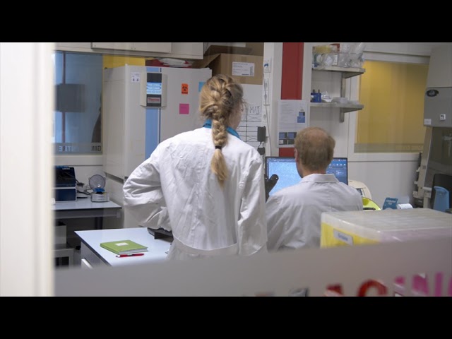
Welcome to the BioImaging Centre
The BIC is a shared ML-I facility for advanced light microscopy at the University of Twente, featuring state-of-the art commercial microscopes, experimental microscopy techniques, image analysis software and ML-II cell- and tissue culture.
BIC Portfolio
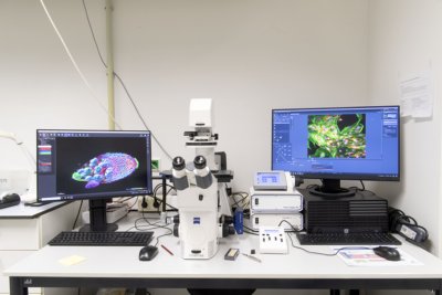
Microscopy
Laser scanning confocal
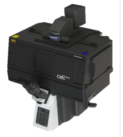
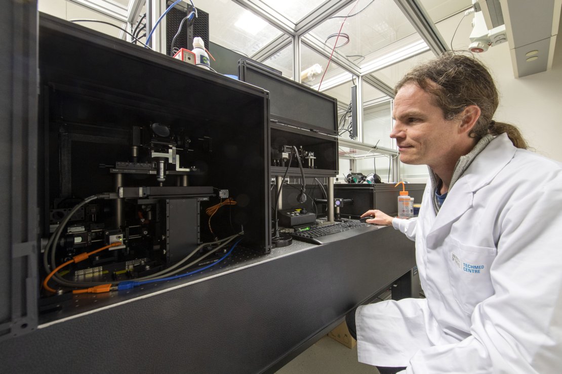
Evident FV4000 + Two-Photon excitation
The Evident FV4000 confocal microscope is a highly advanced, fully motorized inverted microscope from the IX3 series, designed for sophisticated fluorescence imaging in life science research.
Key features
- Confocal
- Resonant galvano scanner
- Laser lines 405, 488, 514, 561, 630 & 730nm
- Two-Photon excitation 750-1300nm
- 6 SilVIR detectors
- Fully motorized
- Incubator
Evident FV4000
The Evident FV4000 confocal microscope is a highly advanced, fully motorized inverted microscope, designed for sophisticated fluorescence imaging in life science research.
Key features
- Confocal
- Laser lines 405, 488, 514, 561 & 630nm
- 2 SilVIR detectors
- Fully motorized
- Incubator
Zeiss LSM880
The Zeiss LSM880 confocal microscope is a powerful imaging tool used in biological and materials science research. It utilizes laser scanning technology to generate high-resolution, three-dimensional images of samples, allowing for the visualization of subcellular structures and fine details of tissues and materials. The LSM880 confocal microscope can be used for a wide range of applications, including fluorescence imaging, live-cell imaging and spectral imaging.
Nikon A1
- Nikon A1
The Nikon A1, our first laser scanning confocal microscope, is a dependable system for fluorescence imaging. It’s not the fastest confocal we have and doesn’t include an incubator for live-cell imaging, but for experiments where these aren’t needed, it’s well-suited. With lower usage compared to other systems, the A1 is readily available for long-term, high-resolution studies, ensuring consistent and accurate results.
Brightfield / Widefield
Zeiss Axio Observer
The Zeiss Axio Observer is a versatile microscope that can perform a range of basic imaging techniques such as brightfield, darkfield, phase contrast, and widefield fluorescence microscopy. This system is ideal for users who want to quickly visualize their samples.
Evident IX83
The Evident IX83 is a fully motorized and automated inverted fluorescence microscope from the IX3 series, designed for advanced life science research. It offers exceptional flexibility and performance for a wide range of imaging applications, including live-cell imaging, time-lapse studies, and multicolor fluorescence.
Echo brightfield (inside laminar flowhood)

Imaging your cells inside a laminar flowhood has never been more easy, with the Echo Rebel. Simply rotate the microscope to use it as an inverted or upright microscope for well plates, microscope slides or other cell/tissue holders.


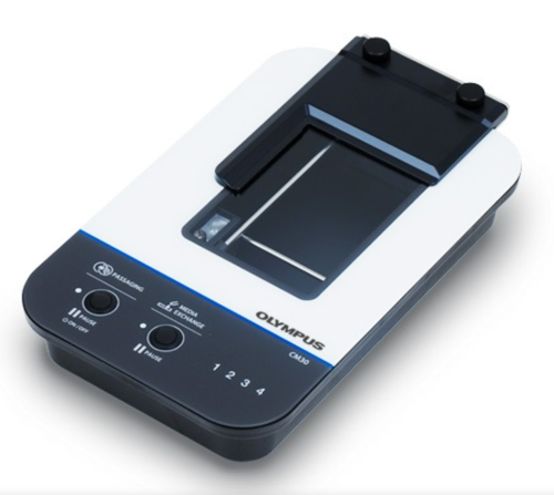
CM30 brightfield (inside incubator)
Remotely monitor, analyze, and share your cell cultures’ health, cell count, and confluency using the reliable quantitative data provided by the automated CM30 incubation monitoring system. The system enables label-free observation, reduces the risk of damage to your cultures, and standardizes your culture workflow.
Holotomography
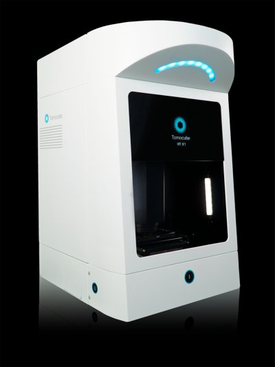
Tomocube
Observations of cellular morphology and activity often rely on labeling target molecules which can be invasive, alter the target molecules, and lack quantitative information. Common imaging tools, such as laser-based microscopes, may also cause phototoxic damage to cells. Holotomography (HT) imaging can visualize cells without labeling by capturing the light scattering properties of cellular materials using low levels of light intensity. The Tomocube HT-X1 Holotomography can collect and selectively color refractive index information to reveal cells and their organelles, while also providing quantitative 3D data such as volume, surface area, and dry mass.
Bioluminescence
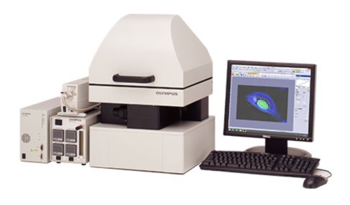
Evident LV200
The LV200 has been carefully designed for long-duration cell bioluminescence. A completely new optical design dramatically increases sensitivity (using the Orca quest 1 for detection) and enables the detailed study of photosensitive cells and luminescence probes at high magnification. The built-in system for temperature control, humidity and gas flow helps to keep the cultured cells or tissue slices in a healthy condition throughout the observation period and the unique light-tight enclosure shields the sample and optics from any external light.
Raman
Witec alpha 300 Raman (532, 633, 785nm)
The WITec alpha300 R is a high-performance confocal Raman imaging microscope designed for non-destructive chemical analysis. It is widely used in materials science, life sciences, and nanotechnology. The alpha300 R is able to use excitation lasers at 532 nm, 633 nm, and 785 nm.
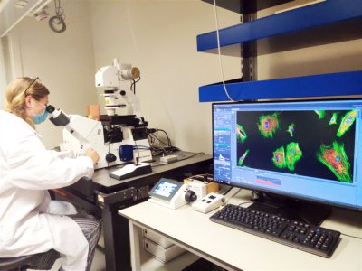
Experimental Microscopy
Two-Photon + wavefrontshaping
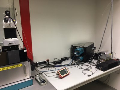
Our homebuilt Two-Photon laser scanning microscope is a powerful imaging tool used in biological and neuroscience research. It uses high-intensity infrared laser pulses to excite fluorescent molecules in living tissues, allowing for the visualization of fine details deep within the sample. Compared to traditional confocal microscopes, it can image deeper and with less photodamage. This microscope is ideal for studying the structure and function of living tissues, including the brain, and organs on a chip systems.
Additionaly our Two-Photon laser scanning microscope includes a wavefront shaping module which can correct for tissue distortions and sample abberations which allows for deeper imaging of biological samples. This module enhances the microscope's capabilities, enabling researchers to visualize previously inaccessible structures and processes within living tissues.
Flowcytometry
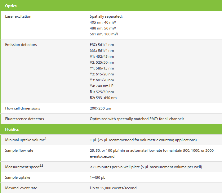
Miltnyi VYB analyzer
MILTENYI VYB ANALYZER
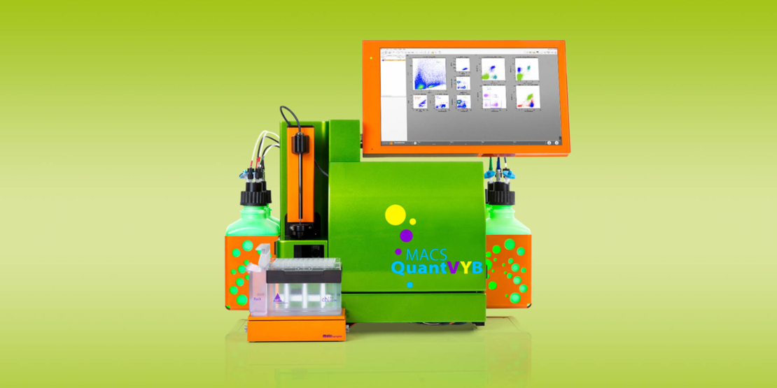
The Miltenyi VYB analyzer is all about performance, automation, compactness, and convenience. It combines all these features, but also comes with a uniquely configured optical bench, featuring violet, yellow, and blue lasers. It is a perfect match for users utilizing fluorescent protein reporters or for users that want to simply use PE and FITC conjugates.
Biological Sample facilities
ML - I
Imaging inside Laminar Flowhood
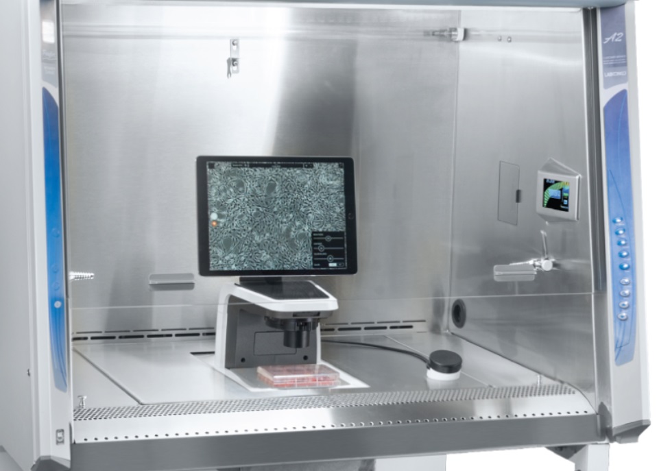
Imaging your cells inside a laminar flowhood has never been more easy, with the Echo Rebel. Simply rotate the microscope to use it as an inverted or upright microscope for well plates, microscope slides or other cell/tissue holders.
Balances / pH-meter / Pipet sets
For handling, transfering, measuring or weighing of your samples you can use the balances, pH-meter, pipet sets, pipet tips, gloves, microscope slides available to every BIC user.
Fume Hood
Fume hood for (chemical) sample handling
ML - II
- CRISPR-Cas9 available on request
- CO2 incubators, laminar flow hoods, centrifuges, freezer
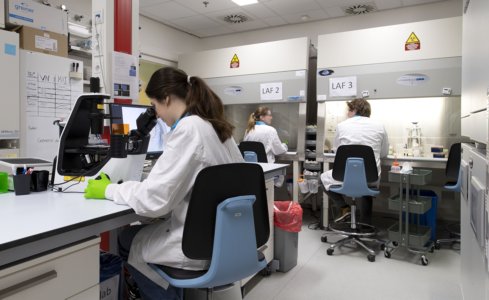
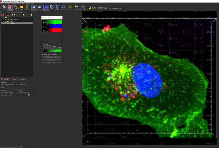
Image analysis software
The BIC presents IMARIS as image analysis software for data analysis free of charge to all of our users. The software can be used on the stand alone Imaris PC in the BIC lab (ZH161) or via Remote Desktop access.
If you want to make use of IMARIS, please send an E-mail to: TNW-BIC-Support@utwente.nl
We also offer help with other image analysis software like ImageJ, FIJI, MicroManager.
Our goal is to assist researchers in addressing complex imaging challenges, providing training and support to users of our facility, to ensure that users have the skills and knowledge needed to operate the equipment effectively.
In addition, our technicians are available to troubleshoot issues and provide guidance on experimental design. For researchers requiring more advanced imaging solutions, our technicians can also work with them to develop customized imaging protocols and techniques.
The BIC is available for both University of Twente users and external users.
HOw to use the BIC facility
In order to request a device introduction and use the BioImagingCentre facility, we kindly ask you to fill in the following ONLINE FORM.
In this way, we have all the necessary information, and we will send you a proposal for a date and time to introduce you to one of our systems and facilities.
The BioImaging Centre is a TechMed facility and affiliated with the NL-BioImaging AM distributed infrastructure.