

The silent epidemic: pelvic floor disorders
Pelvic floor disorders affect an estimated one in three women, yet they often remain under-recognised. Incontinence, prolapse, and pain are not only physically burdensome but also have a significant impact on daily life and self-esteem. Despite this, these conditions receive relatively little attention in research and clinical care. Anique Bellos-Grob aims to change that. From the TechMed Centre at University of Twente, she is committed to improving diagnosis and treatment using advanced medical technologies.
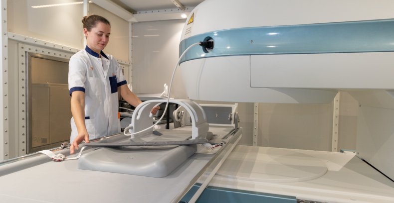
Upright MRI: A revolution in imaging
Traditional MRI scans are performed lying down, which can distort the view of the pelvic floor. “Many symptoms occur while standing or moving,” Bellos-Grob explains. That’s why she uses a unique upright MRI scanner, allowing the body to be examined in a natural posture. This technology is scarcely available in the Netherlands but offers unprecedented possibilities. “We can see how organs shift under pressure—something that’s not always visible when lying down.” These images not only provide new insights but also support more accurate treatment decisions.
Between innovation and implementation
However, the technology also presents challenges. The scanner Bellos-Grob uses is the only one of its kind in the Netherlands and is not intended for routine clinical use. She regularly receives requests from patients and doctors but has to decline. “We’re not a hospital, and without reference images from healthy individuals, we can’t yet determine whether something that looks unusual in a standing position is actually abnormal.” This makes widespread clinical application difficult—for now.
Still, Bellos-Grob remains optimistic. “We’re seeing things we simply couldn’t see before.” The upright MRI paves the way for personalised care tailored to each woman’s unique anatomy. The research also offers opportunities for young scientists and students eager to contribute to more equitable healthcare. “We’re only at the beginning,” she says. “But what we’re learning already could make a real difference for thousands of women. And that’s exactly why this work matters.” Bellos-Grob believes that with proper validation and AI support (see box below), these techniques could fundamentally improve care—especially for the many women currently falling through the cracks.
4D ultrasound: real-time insight into movement
In addition to upright MRI, Bellos-Grob also uses 4D ultrasound. For pelvic floor imaging, a volumetric ultrasound probe is placed externally against the labia. This allows the underlying muscles and structures—such as the sphincter and pelvic floor muscles—to be visualised in real time during contraction and relaxation, without the need for internal examination.
“We can now see exactly how the pelvic floor muscles contract and relax during actions like coughing or straining,” she says. These dynamic images help to better understand functional disorders and enable more targeted treatment. The combination of both techniques—static and dynamic—provides a comprehensive view of the pelvic floor in action.
Although promising, 4D ultrasound is far from straightforward. Interpreting the images requires specialist knowledge, and standardisation is still lacking. Bellos-Grob points to the absence of objective benchmarks: “What constitutes ‘normal’ pelvic floor function? And how do you quantify it?” Moreover, many women struggle to activate the correct muscles; incorrect use of abdominal or gluteal muscles is common. The challenge lies not only in the technology but also in patient interaction and translating findings into usable diagnostics.
how 4D ultrasound aids diagnosis after a severe tear
One of the most severe complications during vaginal birth is a so-called third- or fourth-degree tear, where the external anal sphincter is partially or completely torn. This occurs more often than many realise and can lead to faecal incontinence later in life. Yet in most hospitals, no imaging is performed to assess how well the muscle has healed. With 4D ultrasound, it is possible to accurately determine whether the muscle is still anatomically intact and functioning properly.
“We can even see which part of the muscle no longer moves during contraction,” Bellos-Grob explains. This provides valuable information for follow-up care, such as pelvic physiotherapy or considering a caesarean section for future births. The technique is still under development and requires expert interpretation, but it already offers hope to women who have struggled for years with unexplained symptoms.
personalised care for every woman
Trained as a technical physician, Anique Bellos-Grob has always sought a role that bridges science and clinical practice. That curiosity brought her to the University of Twente, where she now conducts research at the intersection of medicine and technology. She works closely with clinical partners such as surgeons and gynaecologists at ZGT and MST, and actively involves patients in her research. “Their experiences are invaluable. They help us ask the right questions.”
The ultimate goal? Personalised care tailored to each woman’s body and symptoms. “We want to reach a point where imaging can predict which treatment will work best,” says Bellos-Grob. That requires smart algorithms, large datasets, and interdisciplinary collaboration. At the TechMed Centre, that cross-pollination is thriving. “We’re bringing together engineers, clinicians, and data scientists to shape the healthcare of tomorrow.”
For students eager to make an impact in healthcare, now is the time to get involved. “Women’s health has long been overlooked, but that’s finally changing,” says Bellos-Grob. She hopes that young researchers will be drawn to both the societal relevance and the technological challenge. “At the University of Twente, you have the opportunity to truly make a difference – for patients, for science, and for yourself.”