
PhD Student
E-mail: b.liszka@utwente.nl
Telephone: +31 53 489 4225
Fax: +31 53 489 3511
Address: Faculty of Science and Technology
Medical Cell BioPhysics (MCBP)
Building: Carré
Room: CR4435
Drienerlolaan 5
P.O. Box 217
7500 AE Enschede
The Netherlands
Project goals
Understanding mechanism of crystals formation and their growth at organic surfaces in early stage. Developing methods which enable to detect nano-crystal and follow their growth.
Project motivation
Water treatment is necessary in many industrial scale processes, such as: waste water treatment, drinking water preparation, industrial installation. Mineral components are ubiquitous in water and may at intermediate stages during water treatment, lead to mineralization. Membranes, made from organic polymers, are often used in filter systems. The membrane materials are sensitive to fouling, which adversely affects quality. The resulting limited lifetime of membrane systems is undesirable. Method to understand and detect early (nano)crystal formation may help to protect surfaces.
Methods
Mineralization solutions and organic surfaces on which crystals growth occurs are study by application of various techniques such as : Raman Micro Spectroscopy, SEM, microfluidics platforms, pH sensors system for kinetic measurements and combination of those methods.
Microfluidics is a technique which enables to miniaturize macro process to micro scale. In such a device conditions occurring during water treatment can be mimic under controlled and defined parameters. Integration of a microfluidics platform with a Micro Raman Spectroscopy enables for physico-chemical analysis of formed minerals on a surfaces. The method offers high resolution which is governed by light wavelength and it is order 400 nm. The sensitivity is also, high enough to detect nano-particles. Integration of both techniques allows for in situ high resolution imaging of processes under ambient conditions. Due to all of this aspects, Raman-Opto-Fluidic becomes an attractive toll not only to study membrane scaling but also, in fields such as :medicine, clinical diagnostic and biotechnology.
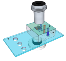
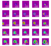
Figure 1. Left Schematic representation of microfluidic chip. Right Raman cluster images acquired in 4h showing single crystal growth at polymer membrane.
The system of multiple pH sensors enables to study kinetic of calcium carbonate mineralization in solution. The changes of pH reflect stages of crystals formation such us: nucleation, growth and ageing. The scanning electron microscopy (SEM) enables to study detailed structure of crystals, detect nano-sized particles and quantify them.
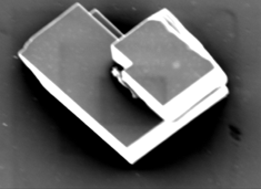


Figure 2. Left SEM image of calcium carbonate crystal -calcite. Right Characteristic Raman spectrum of calcite.
Potential application of methods: Raman –Opto- Fluidic, SEM-Raman is a sensor for early scaling detection on a membrane.