The manuscript ‘Fluorescence based Quantification of Gastrointestinal Perfusion: A step towards an Automated Approach’ by Harry Vaassen, Bryan Wermelink, Bob Geelkerken and Daan Lips is accepted in the Journal of Laparoendoscopic & Advanced Surgical Techniques.
Inadequate local or systemic bowel perfusion is regarded as a substantial factor in complications after gastrointestinal surgery. In current practice, blood supply is assessed peroperatively based on clinical observations, but this lacks accuracy and objectivity. Fluorescence angiography (FA) has emerged in recent years as a promising tool for yielding intraoperative insights in local perfusion dynamics. It involves the injection of indocyanine green (ICG) and monitoring of its fluorescent appearance in tissue.
Currently, FA data is mostly evaluated in a qualitative fashion, making it sensible to misinterpretation and subjectivity. Literature shows promising predictive value of quantified parameters based on ICG inflow. Quantification of FA can be achieved by measuring the intensity over time in a specific region of interest. However, there is no general consensus on the exact acquisition and interpretation of these parameters.
In this work we showed that an automated changepoint detection algorithm is able to accurately and consistently extract inflow-based parameters from FA videos. Moreover, we investigated the characteristics of parameter T_1/2, defined as the time between first intensity increase and the point of reaching 50% of the maximal intensity, in patients with normal intestinal perfusion. The automatic algorithm also allowed the creation of immersive ‘cartograms’, based on inflow-parameters.
We consider this study as the foundation on which a reliable and objective perfusion evaluation system can be built. Future studies will include patients with impaired bowel perfusion and pursue threshold values to identify insufficient intestinal blood supply.
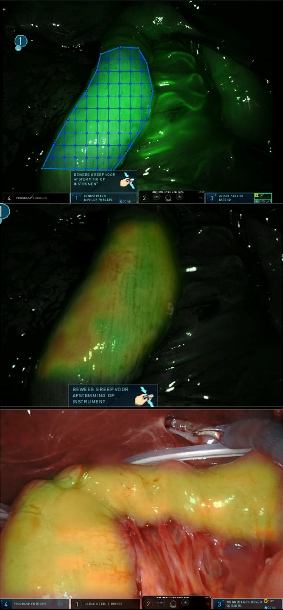
a) Manual segmentation and partitioning of small intestine in FA recording. |


