Tunable blood oxygenation in the vascular anatomy of a semi-anthropomorphic photoacoustic breast phantom by Maura Dantuma, Saskia Kruitwagen, Javier Ortega Julia, Rutger van Meerdervoort and Srirang Manohar was accepted in the journal of biomedical optics.
Recovering accurate oxygenation estimations in the breast with quantitative photoacoustic tomography (QPAT) is not straightforward. Accurate light fluence models are required, but the unknown ground truth of the breast makes it difficult to validate them. Phantoms are often used for the validation, but most reported phantoms have a simple architecture. Fluence models developed in these simplistic objects are not accurate for application on the complex tissues of the breast.
We present a sophisticated breast phantom platform for photoacoustic (PA) and ultrasound (US) imaging in general, and specifically for QPAT. The breast phantom is semi-anthropomorphic in distribution of optical and acoustic properties and contains wall-less channels with blood.
3D printing approaches are used to develop the solid 3D breast phantom from custom polyvinyl chloride plastisol (PVCP) formulations and additives for replicating the tissue optical and acoustic properties. A flow circuit was developed to flush the channels with bovine blood with a controlled oxygen saturation level. To showcase the phantom’s functionality, PA measurements were performed on the phantom with two oxygenation levels. Image reconstructions with and without fluence compensation from Monte Carlo simulations were analyzed for the accuracy of oxygen saturation estimations.
We present design aspects of the phantom, demonstrate how it is developed, and present its breast-like appearance in PA and US imaging. The oxygen saturations were estimated in two regions of interest with and without using the fluence models. The fluence compensation positively influenced the SO2 estimations in all cases and confirmed that highly accurate fluence models are required to minimize estimation errors.
This phantom allows studies to be performed in PA in carefully controlled laboratory settings to validate approaches to recover both qualitative and quantitative features sought after in in-vivo studies. We believe that testing with phantoms of this complexity can streamline the transition of new PA technologies from the laboratory to studies in the clinic.
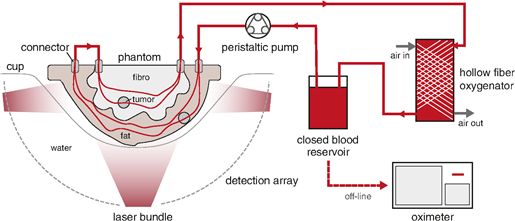
Figure 1: The setup used during the PA measurements. The tunable oxygenation breast phantom is placed in a cup inside the Twente PAM 2 system. A peristaltic pump drives the blood from the reservoir through the circuit, where blood is being oxygenated by a hollow fiber oxygenator. Air flow through this oxygenator can be controlled to regulate the blood oxygenation. Sodium hydrosulfite can be added to the blood reservoir to deoxygenate the blood. Samples are withdrawn from the oxygenator and the oxygenation is measured with the oximeter.

Figure 2: (a) LMIPs of the medial view of the PA measurements with 755 and 1064 nm for the two blood oxygenation levels. (b) The average reconstructed pressures from the vessel pixels in the ROIs highlighted in (a) together with the blood absorption coefficients for the respective wavelengths and oxygen saturations.


