K. Roya-Kouchaki 1, H.L. Offerhaus 1, G. Muñoz González,2 A. Luchicchi2
1 Optical Sciences, UTwente 2 Amstardam UMC, Dept. Anatomy and Neurosciences
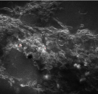
The onset of Multiple Sclerosis (MS) has traditionally been sought in the immune system (outside-in-model). More recently it has been proposed that MS originates inside the brain and the immune system is antagonized later [1]. We use Coherent anti-Stokes Raman Scattering (CARS) microscopy to detect the start of demyelination. We cannot resolve the distance between myelin layers (20-100nm) but try to capture the vibrational spectrum of the water in between the layers. This spectrum changes when the layers widen and the water molecules form more hydrogen bridges. We will present the first exploratory measurements in this region.
[1] “Axon-Myelin Unit Blistering as Early Event in MS Normal Appearing White Matter” A.Luchicchi et al., ANN NEUROL 2021;00:1–15, DOI: 10.1002/ana.26014