- Wooje Lee - PhD student
- Herman Offerhaus - Chair
This project is part of a bigger project: New technology for monitoring CANCER therapy through extracellular vesicle Identity (CANCER-ID)
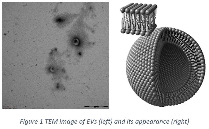
Tumor-derived extracellular vesicles (tdEVs) may contain information important for the management of cancer patients. Their presence in biofluids of cancer patients renders them a promising biomarker for the diagnosis of cancer, the monitoring of therapy and the profiling of the tumor leading to a therapy tailored to the individual. Within the framework of the Cancer-ID program, novel techniques for the detection, isolation and characterization of tdEVs are being developed. EVs of different origins have been prepared and are being characterized with Raman spectroscopy, fluorescence microscopy and electrical impedance spectroscopy. Functionalized microfluidics and microsieves approaches are being developed to isolate them specifically from heterogeneous patient samples.
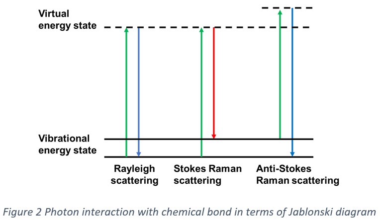
To reveal chemical information of EVs we use spontaneous Raman spectroscopy which is a type of vibrational spectroscopy technique based on inelastic scattering by molecules. When incident photons are scattered by molecules, some are scattered with particular energy shifts, a phenomenon called Raman scattering. Raman microscopy is used to investigate structural and compositional information of a specimen. Since the optical technique yields the fingerprint of chemicals, it has been widely used in biological and pharmaceutical fields. It has been applied to identify differences in tissues and cells. Convincing spectral differences have been demonstrated between cancer cells and healthy cells, based on lipid droplet content, carotenoids and ratio between different proteins.