Cells from different origins leave behind extracellular vesicles (EVs) in blood. Patients with cancer have tumour cells that do the same, but these tumour-derived extracellular vesicles (tdEVs) are vastly outnumbered by lipoproteins and EVs from other cells such as red blood cells, platelets and leukocytes. Researchers of the University of Twente and the University of Amsterdam managed to trap and accurately detect single tdEVs with a laser. The researchers present their results in the prestigious Journal of Extracellular Vesicles.
Detecting without labels
In cancer diagnostics, extracellular vesicles attract a lot of attention. The nano-sized particles are naturally released from all types of cells. When you detect tumour-derived extracellular vesicles in a patient’s blood, they can inform you about the effectiveness of a cancer treatment. The problem is that the number of tdEVs in a patient’s blood is extremely small, compared to lipoproteins and other ‘normal’ EVs. “Distinguishing tdEVs from other particles in plasma is challenging even with labels such as antibodies, which are not specific enough and can clump together and create a false tdEV”, says UT researcher Agustin Enciso-Martinez. His method can detect tdEVs with an accuracy of over 95 percent in a label-free manner.
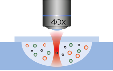
Figure 1: Trapping particles with a laser
Unique fingerprint
The use of light to trap tiny particles earned Arthur Ashkin the Nobel Prize in Physics in 2018. This principle of optical trapping has been used by UT researchers to develop a method to trap and accurately detect single tdEVs in suspension using a laser. The light that bounces off a trapped particle can be measured to know that you have successfully trapped a particle. When the light bounces of a particle, the colour changes ever so slightly according to the properties of the molecules inside. This change can also be measured and gives tdEVs a unique fingerprint which can be used to tell them apart from other particles in blood plasma.
More research is needed before this technique can be reliably used in the clinic. Enciso-Martinez says: “We showed the unique fingerprint of tdEVs in test suspensions, and currently we are testing our method on blood from cancer patients.” Blood has a vast number of other particles with a very similar size as tdEVs which makes identification harder. Also, the research was done with vesicles from prostate cancer, other tdEVs remain to be tested.
About the paper
The research is part of the Cancer-ID programme, which is co-funded by the Netherlands Organization for Scientific Research (NWO). The paper ‘Label-free identification and chemical characterisation of single extracellular vesicles and lipoproteins by synchronous Rayleigh and Raman scattering’, by Agustin Enciso-Martinez (UT), Edwin van der Pol (UvA), Chi M. Hau (UvA), Rienk Nieuwland (UvA), Ton G. van Leeuwen (UvA), Leon W.M.M. Terstappen (UT) and Cees Otto (UT) is published online in the Journal of Extracellular Vesicles.


