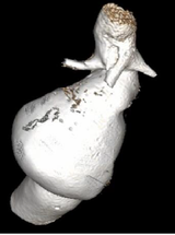
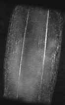
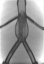
Vascular pathologies are often well imaged using MRI because of its variable contrast mechanisms (contrast agent enhanced, flow enhanced, black blood, white blood, etc.) and possibility to image large FOVs in 3D, as opposed to ultrasound. A disadvantage compared to other imaging modalities is mainly found in the total imaging time. Also in this research upright imaging might give a different perspective on the anatomy. For example, it is well known that the diameter of veins is greatly influenced by body posture. Next to that, therapies of the vasculature often involve placing supporting structures, so-called endografts, within the vessels. One often occurring problem with these endografts is leakage; the hypothesis of our clinical partners is that this might also be caused by posture.
With this subject the research questions we try to answer are: “Can upright MR imaging of the venous system give us valuable clinical information about pathologies?” and “Can the quality of endografts be assessed in upright position?”. There are several research goals in this line. First the venous system is assessed in different areas such as in the head and neck region to assess brain outflow and venous pressure. Additionally venous insufficiency can be studied in the legs. It has already been shown that venous imaging can be performed and their behavior is dependent of body posture but the clinical relevance is still missing.
Secondly, in the endograft study the first steps were made to see whether the endografts could be imaged robustly in vivo using our low-field MRI scanner . The next goal is to see whether we can define reliable measures to define their effectiveness, for example the total height of which they are fully connected to the arterial wall. The final goal here would be to detect of leakages in upright position that were up until now without a known cause.
This research is the main focus of the PhD project of Jordy van Zandwijk. We have a collaboration with a large consortium of vascular surgeons of which prof. Bob Geelkerken (M3I/MST) and prof. Jean-Paul de Vries (M3I/UMCG) are our main contacts.
|
|
|