Laser Speckle contrast imaging and Photoacoustic imaging in PAOD patients in the hybrid Operating room
In peripheral arterial occlusive disease (PAOD) one or more arterial stenosis, cause a change in haemodynamics. The change in haemodynamics results is an insufficient blood supply in the legs. In end stadium PAOD this can result in severe claudication or tissue loss of the affected tissue. To prevent tissue loss or to remedy severe claudication, PAOD patients are treated endovascularly by means of Percutaneous transluminal Angioplasty (PTA), possibly supplemented with the placement of a stent. Since January 2016, these endovascular procedures, in MST Enschede, are carried out in the Hybrid Operating Theatre (HOT). In the HOT, visualization of blood and oxygen delivery to the affected tissue, the microcirculation, is not yet possible. Therefore, besides the conventional fluoroscopy and angiography, a second imaging module should be present in the PAOD endovascular operation program, that can visualise the microcirculation of the foot. Imaging methods that can visualise the microcirculation are laser Speckle contrast imaging (LSCI) and Photoacoustic imaging (PAI). The goal of the current project is to evaluate the added clinical value of several microcirculation techniques in a HOT setting.
Laser Speckle Contrast Imaging
LSCI is a contactless optical technique generating microvascular blood flow maps with high temporal and spatial resolution. Interference patterns, formed by the light scattered from the tissue, are recorded with a digital charge-coupled-device (CCD) camera. Movement of red blood cells in the microcirculation changes the interference pattern in time. This change leads to blurring of the recorded laser Speckle image (the higher the velocity of the red blood cells, the more blurred the image). By analysing this blurring, blood flow maps are generated and displayed instantaneously. The video below shows an example of post occlusive reactive hyperemia, imaged with LSCI.
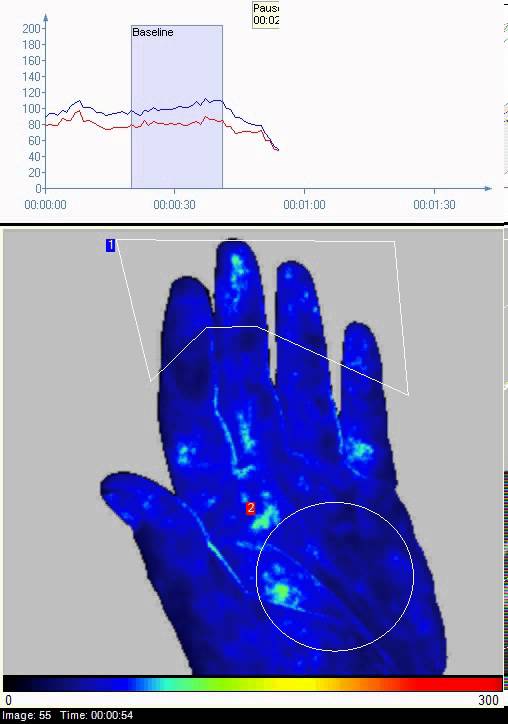
Source: Perimed AB1
Photoacoustic Imaging
In PAI, a short pulse of light is emitted. This light is absorbed by tissue chromophores. A chromophore is the part of a molecule that is responsible for its colour. Absorption of light by chromophores create a local instantaneous temperature rise. The volume containing the chromophores instantaneously expands and builds up a pressure. This pressure build up leads to the emission of an ultrasound wave. This wave can be detected with an ultrasound transducer array at the tissue surface. PAI combines high penetration depth and submillimetre ultrasound resolution. PAI can be used for visualizing the presence of small blood vessels, the content of haemoglobin and its degree of oxygenation.
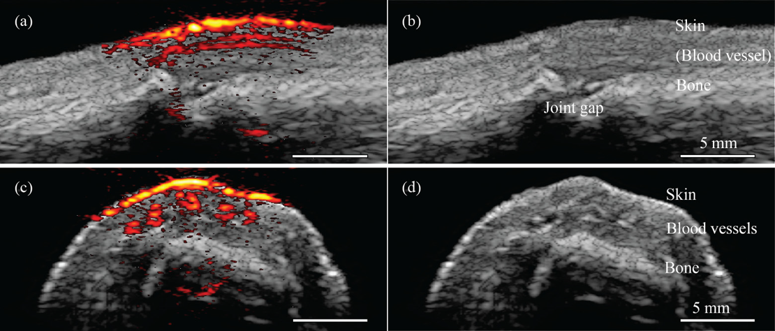
Figure 3: PAI image combined with ultrasound of a human proximal interphalangeal joint in sagittal (a) and transverse (b) planes, with the upper part of the image corresponding to the dorsal side of the finger. On the right side, (b) and (d) show corresponding ultrasound only with anatomical structures indicated2.
The goal of this pilot study is to determine if LSCI and/or PAI could be used for the visualisation of the microcirculation during a PAOD procedure in a HOT setting. We will investigate the stability and reproducibility of LSCI and PAI when applied in the HOT. If one or both techniques show high stability and reproducibility, the outcome parameters of the techniques need to be correlated to the clinical outcome after the endovascular procedure.
References
Images:
1. Perimed. Perimed Laser Speckle Contrast Imaging [Internet]. Available from: http://www.perimed-instruments.com/products/pericam-psi
2. Daoudi K, van den Berg PJ, Rabot O, Kohl A, Tisserand S, Brands P, et al. Handheld probe integrating laser diode and ultrasound transducer array for ultrasound/photoacoustic dual modality imaging. Opt Express [Internet]. 2014;22(21):26365. Available from: https://www.osapublishing.org/oe/abstract.cfm?uri=oe-22-21-26365
LSCI:
Ruaro B, Sulli A, Smith V, Paolino S, Pizzorni C, Cutolo M. Short-term follow-up of digital ulcers by laser speckle contrast analysis in systemic sclerosis patients. Microvasc Res [Internet]. Elsevier Inc.; 2015;101:82–5. Available from: http://dx.doi.org/10.1016/j.mvr.2015.06.009
PAI:
Daoudi K, van den Berg PJ, Rabot O, Kohl A, Tisserand S, Brands P, et al. Handheld probe integrating laser diode and ultrasound transducer array for ultrasound/photoacoustic dual modality imaging. Opt Express [Internet]. 2014;22(21):26365. Available from: https://www.osapublishing.org/oe/abstract.cfm?uri=oe-22-21-26365