Dedicated breast CT
Dedicated breast computed tomography is a novel imaging technology being developed for breast cancer imaging. It provides true 3D breast imaging at low doses with high spatial resolution, therefore overcoming the limitation of tissue superposition in mammography and digital breast tomosynthesis. Potential applications are in various stages of the breast cancer pathway, including diagnosis, staging, treatment planning, and therapy response monitoring.
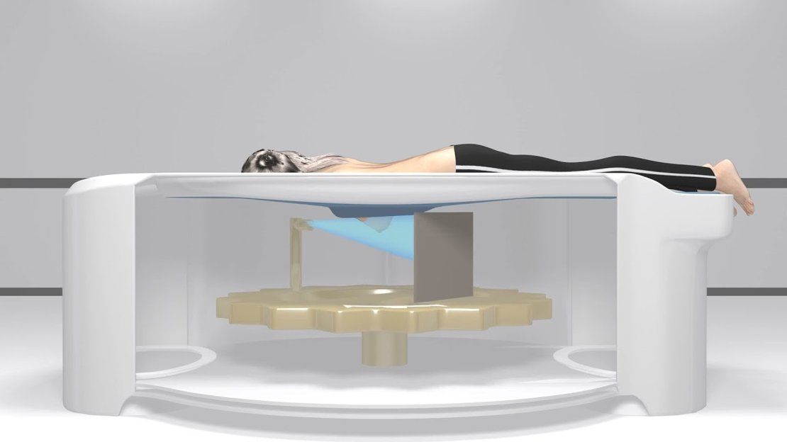 Figure 1: The breast CT system (Image source and copyright: Koning Health)
Developments to improve image quality are ongoing in multiple parts of the imaging pipeline, including in image acquisition and reconstruction, as well as image processing and analysis. In the BREAST4D project, dedicated breast CT is used to image the wash-in and wash-out of iodinated contrast agent in the breast. This will enable the assessment of functional information of the breast and its abnormalities by quantitatively evaluating the iodine in and outflow. This information could be beneficial in staging, treatment planning, and monitoring of breast cancer.
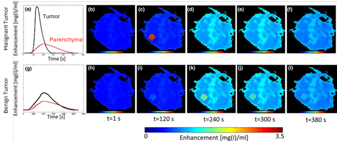 Figure 2: 4D breast CT expected results showing differences in contrast enhancement curves between a malignant (top) and benign (bottom) tumor model. Figure adapted from Caballo, Marco, Ritse Mann, and Ioannis Sechopoulos. "Patient‐based 4D digital breast phantom for perfusion contrast-enhanced breast CT imaging.” Medical physics 45.10 (2018): 4448-4460.
PROJECT GOAL
The goal of this research line is to further develop the breast CT system hardware and software, using new X-ray sources or types of detectors, to improve image quality and minimize exposure to ionizing radiation, and to develop methods for accurate 4D breast CT perfusion imaging.
COLLABORATIONS
- University of Twente
- RadboudUMC
CONTACT


