These projects aim to contribute to the validation, calibration, and evaluation of novel quantitative perfusion imaging techniques via phantom modeling. Phantom studies can provide controlled, simplified, and reproducible environments to simulate the circulation and perfusion in different body parts. This allows for the objective and quantitative evaluation of performance of imaging technologies undergoing development or optimization.
PROJECTS
Myocardial perfusion imaging
Perfusion imaging of the brain
Dynamic CT perfusion imaging of the breast
Myocardial perfusion imaging
PROJECT GOAL
The project aims to contribute to the validation, calibration and evaluation of quantitative myocardial perfusion imaging via phantom modeling.
MOTIVATION
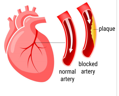
Plaque buildup inside the coronary arteries (arteries that supply the heart muscle) can cause a flow limitation towards the myocardial tissue (heart muscle tissue). If left untreated, this tissue could eventually die, leading to infarction. Proper medical decision-making is key, since coronary artery disease remains one of the most common causes of death in the western world.
Myocardial perfusion imaging is an important imaging technique to diagnose perfusion / blood flow defects. During such diagnostic test, a radio-active tracer is injected into a patient’s blood stream. Then the distribution of tracer inside the heart muscle is imaged over time using positron emission tomography (PET) or single photon computed tomography (SPECT) imaging. A reduced uptake in a certain region (relative to other regions) indicates a perfusion defect. However, evaluation of the resulting images is rather subjective and could underestimate a patient’s risk.
Blood flow quantification software, based on indicator dilution theory, could improve diagnostic accuracy and precision. Such software is tested extensively for PET imaging. However, can we also use it for clinical practice with SPECT scanners?
COLLABORATIONS
- Multi Modality Medical Imaging group (UT)
- Ziekenhuis Groep Twente hospital
- European Institute for Molecular Imaging Münster
Perfusion imaging of the brain
Project goal
To contribute to the validation, calibration, and evaluation of quantitative cerebral perfusion imaging via phantom modeling.
Motivation
“Time is brain”
This commonly-used phrase emphasizes that in the case of a stroke, every second counts; decrease in brain perfusion may cause irreversible brain damage within minutes. Reducing the time between diagnosis and intervention could save millions of neurons. Currently, computed tomography perfusion (CTP) or perfusion-weighted magnetic resonance imaging (PW MRI) are used to determine the affected brain tissue before intervention is initiated. A possibility to save time could be to image and evaluate brain perfusion at the interventional suite, therefore eliminating the extra stop for diagnostic imaging with CTP or PW MRI. In this way, revascularization can also be evaluated during intervention, preventing the need for future re-interventions.
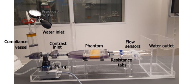
A previous study has shown that cone-beam CT (CBCT) C-arm systems could be used if its temporal and spatial resolution is improved, and image noise is reduced. If this is achieved, imaging obtained with a biplane flat panel angiographic system is suitable to establish clinically usable perfusion maps. To investigate and optimize the use of CBCT for diagnosis, evaluation, and treatment of ischemic stroke, a comparison needs to be made between the current imaging modality, CTP, and the perfusion maps obtained with CBCT. For this, a dynamic perfusion brain phantom could be used to study cerebral perfusion imaging in CTP and CBCT and the two modalities compared.
Figure 2 shows the phantom set-up which is used to study cerebral perfusion imaging in CT.
Collaborations
· Multi-Modality Medical Imaging group (UT)
· Universitair Medisch Centrum Groningen (UMCG)
Dynamic CT perfusion imaging of the breast
Project goal
To contribute to the validation, calibration, and evaluation of quantitative breast dynamic CT perfusion imaging for treatment planning and response monitoring and prediction.
Motivation
Dynamic CT perfusion (dCTP) imaging of the breast, BREAST4D, has the potential to improve treatment planning, response monitoring, and outcome prediction in patients with breast cancer. The dCTP scans will consist of constantly acquiring CT images of the breast throughout the period that the iodinated contrast flows in and out of the tumor, resulting in a 4D CT image of the breast and the tumor perfusion process. These images will be analyzed using radiomics and AI methods.
As part of this project, the quantitative accuracy of the dCTP images needs to be validated against a known truth. Therefore, we aim to develop a dynamic breast phantom in which the iodinated blood flow and perfusion properties of the breast can be simulated.
Collaborations
· Multi-Modality Medical Imaging group (UT)
· Radboud University Medical Center
CONTACT

