The PAMMOTH project and 3rd Generation Imager
PROJECT ACRONYM PAM3
FUNDED BY European Horizon 2020 PAMMOTH project under grant agreement No 732411, an initiative of the Photonics Public Private Partnership.
PRINCIPAL RESEARCHERS Maura Dantuma; David Thompson; Saskia Kruitwagen, Rianne Bulthuis
SUPPORT Johan van Hespen
SUPERVISOR Srirang Manohar
COLLABORATION PA Imaging R&D BV, University College London (UCL), Brno University of Technology (BUT), Medisch Spectrum Twente (MST)
BACKGROUND [1] With Europe’s aging population, its high incidence of breast cancer, its tightening health-care budget, and room for improvement in conventional breast imaging modalities, there is a need for a technique that can provide images with high specificity, contrast and spatial resolution. Photoacoustic imaging may turn out to be that technique. In photoacoustics, the contrast is dependent on light absorption in tissue. Since blood, fat and water in tissue absorb light depending on the wavelength used, the method can provide spectroscopic (molecular) specificity to these and other tissue constituents. Blood is of interest as a biomarker since more blood vessels are often present at tumor sites.
OBJECTIVE PAMMOTH brought together experts from various disciplines to work on a new generation system for imaging the breast using both photoacoustics and ultrasound. Academia, industry and the clinic from 6 different countries were part of the PAMMOTH consortium. The consortium’s objective was to develop, validate and begin exploitation of a dedicated breast imaging device. The proposed device would combine non-invasive 3D photoacoustic imaging with ultrasound imaging to provide full-breast, multimodal images to the radiologist. The device has high through-put, possesses no carcinogenic potential, uses no contrast agent and causes no pain or discomfort to patients.
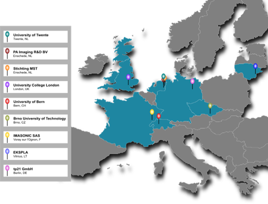
Figure 1 The PAMMOTH partners.
PROJECT DETAILS
System Overview At the conclusion of the project, we have developed the sophisticated PAMMOTH imager. From the photoacoustic mode it is capable of visualizing blood vessels in the breast at superior fields-of-view and depths compared to the state-of-the-art. The ultrasound mode which at this point recovers only the sound speed (and not yet ultrasound reflectivity) provides two valuable inputs: i) the heterogeneous distribution of speed of sound in the breast necessary for an accurate PA reconstruction, ii) insights into tissue-type distribution in the breast. This latter information shows potential for identifying tumors based on speed of sound contrast.
The following highlights can be noted:
- The clinical prototype PAMMOTH, boasting sub-systems that are technically beyond the state-of-the-art, has been developed, tested and is being used to image patients in a clinical study.
- The developed ns laser has among the highest energy outputs per ns pulse. The OPO pumped by the laser allows fast tuning and high efficiency. The light is coupled to the PAMMOTH imager using an optical fiber bundle, designed to accept high energy pulses, split in 40 outputs.
- The 512 ultrasound transducers arranged in hemispherical geometry provide a low minimum detectable pressure with a wide fractional frequency bandwidth.
- The 512 multi-channel Data-Acquisition System is also equipped to transmit ultrasound from the same transducers for ultrasound imaging.
- Cups of various sizes to immobilize the breast during measurements have been produced to hold the breast in the imaging bowl.
- A novel algorithm has been developed for the specific task of reconstructing intermediate images on the fly as projections are being acquired.
- Novel algorithms for speed of sound reconstruction from ultrasound data have been developed and tested.
- A photoacoustic image reconstruction (using a priori known speed of sound map) based on an iterative approach has been developed.
- The algorithm for quantitative photoacoustic reconstruction has been developed.
- A suite of test and calibration objects has been developed.
In vivo Study A total of 8 healthy volunteers have been imaged. See Figure 2 for an example. The clinical study is on hold due to upgrade of the system.
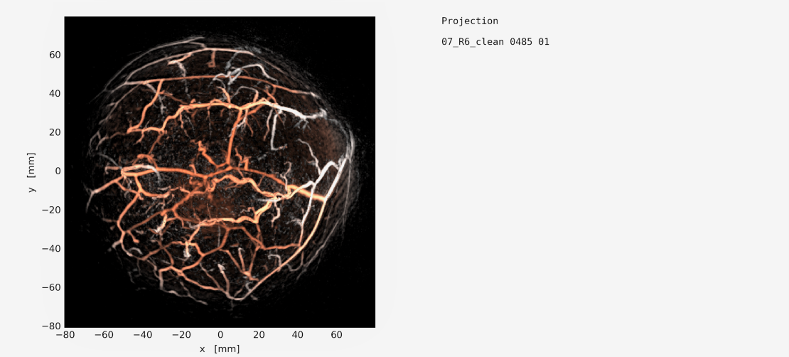
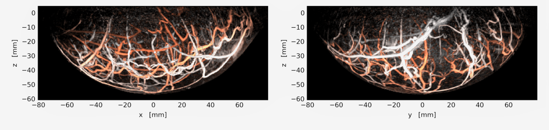 Figure 2: Photoacoustic image of the breast acquired with hemispherical detection geometry. Image courtesy: PA Imaging R&D BV. (Manuscript in peparation.)
OPPORTUNITES
We need highly creative, industrious and passionate students to help us move the field further. The backgrounds required are in Biomedical Engineering, Applied Physics, Technical Medicine, and Computer Science. If you are interested in making an important contribution to this area, contact the following:


PUBLICATIONS
- PAMMOTH Consortium coordinated by Srirang Manohar: https://www.pammoth-2020.eu/
- Bosschaart, N., & Manohar, S. (2022). New directions for optical breast imaging and sensing: multimodal cancer imaging and lactation research. Current Opinion in Biomedical Engineering, 100380. https://doi.org/10.1016/j.cobme.2022.100380
- Thompson, D., Nagel, J. R., Gasteau, D. B., & Manohar, S. (2022). Laser-induced ultrasound transmitters for large-volume ultrasound tomography. Photoacoustics, 25, 100312. https://doi.org/10.1016/j.pacs.2021.100312
- Dantuma, M. M., Kruitwagen, S. C., Weggemans, M. J., Op't Root, T. J., & Manohar, S. S. (2021). Suite of 3D test objects for performance assessment of hybrid photoacoustic-ultrasound breast imaging systems. Journal of biomedical optics, 27(7), 074709. https://doi.org/10.1117/1.JBO.27.7.074709
- Voets, M. M., Groothuis-Oudshoorn, C. G., Veneklaas, L. H., Manohar, S., Brinkhuis, M., Veltman, J., ... & Siesling, S. (2021). Diagnostics in Patients Suspect for Breast Cancer in The Netherlands. Current Oncology, 28(6), 4998-5008. https://doi.org/10.3390/curroncol28060419
- Dantuma, M., Kruitwagen, S., Julia, J. O., van Meerdervoort, R. P. P., & Manohar, S. (2021). Tunable blood oxygenation in the vascular anatomy of a semi-anthropomorphic photoacoustic breast phantom. Journal of biomedical optics, 26(3), 036003. https://doi.org/10.1117/1.JBO.26.3.036003
- Thompson, D., Gasteau, D., & Manohar, S. (2020). Spatially compounded plane wave imaging using a laser-induced ultrasound source. Photoacoustics, 18, 100154.https://doi.org/10.1016/j.pacs.2019.100154
- Manohar, S., & Dantuma, M. (2019). Current and future trends in photoacoustic breast imaging. Photoacoustics, 16, 100134. https://doi.org/10.1016/j.pacs.2019.04.004
- Dantuma, M., van Dommelen, R., & Manohar, S. (2019). Semi-anthropomorphic photoacoustic breast phantom. Biomedical optics express, 10(11), 5921-5939.https://doi.org/10.1364/BOE.10.005921