Previous meetings 2017 | |||
Date | Speaker(s) | Affiliation | Subject |
July & Augustus | Summer holidays |
|
|
28 June | Ruud van Kaam | Clinical fellow Technical Medicine | Exploring Cortical Spreading Depression and Remote Network Change in Acute Ischaemic Stroke |
31 May | Sofie Carrette | Ghent University Hospital in Ghent, Belgium | Transcranial magnetic stimulation in epilepsy |
17 May | Manu Kalia | Applied Analysis | Extracellular dynamics in a single cell model for cytotoxic edema |
3 May | Boudewijn Sleutjes & Hessel Franssen | University Medical Center Utrecht, Utrecht | Measuring and modelling human peripheral nerve excitability |
5 April | Stephan van Gils | Applied Analysis | Cross-Scale Effects of Feedforward Inhibition during Human Neocortical Seizure Activity |
22 March | Pauly Ossenblok | Kempenhaeghe | Multimodal windows on spontaneous brain activity in epilepsy research |
8 March | Joost le Feber | Clinical Neurophysiology | Loss and recovery of connectivity in an in vitro model of the ischemic penumbra |
25 January | Nikita van der Vinne | Research Institute Brainclinics & Utrecht University | Frontal alpha asymmetry in depression: fact or fiction? A meta-analysis |
11 January | Ida Nissen | VU University Medical Center, Amsterdam | Localizing the epileptogenic zone in interictal MEG recordings using network theory |
Introduction: Acute ischemic stroke is a major cause of death and disability, worldwide. The only treatments of proven benefit are intravenous and intra-arterial recanalization techniques. Treatments to prevent secondary brain damage or promote cerebral recovery have a large potential to improve neurological recovery. However, the pathophysiology of secondary brain damage and recovery are largely unclear and treatments to promote recovery of ischemic brain damage are lacking. With the current study, we aim to obtain insight in potential causes of secondary deterioration, focussing on detection of cortical spreading depression (CSD). In addition, the association between remote network changes in the contralesional hemisphere and functional recovery are studied. For this purpose, we used full band continuous EEG recording within the first two days in patients with acute cortical brain infarcts. Methods: EEG registration was started within 48 hours after symptom onset in patients with acute ischaemic cortical stroke and a National Institutes of Health Stroke Scale (NIHSS) score of ≥ 4. Analysis of CSD was largely done visually. The characteristics of CSD; a large slow potential change (SPC, << 0.1 Hz) accompanied by a simultaneous amplitude depression of spontaneous neuronal activity (> 1 Hz), a spread of SPC and depression and unilateral presentation in the lesioned hemisphere, were sought for in the raw time series in epochs of one hour. For analysis of remote network changes in the contralesional hemisphere, delta-alfa ratio (DAR), magnitude squared coherence (MSC), and weighted phase lag index (WPLI) were calculated from the ipsilateral and contralateral hemispheres and were compared to a healthy control group. Results: Eighteen patients were included in the study. Visual inspection revealed many artefacts and a poor signal to noise ratio, to be largely traced to muscle contraction and movement artefacts. No single characteristics of CSDs was found in the data. Analysis of remote network disturbances showed increased DAR (median: 2.43, IQR 2.01) in the contralateral hemisphere, compared to the control group (median: 0.47, IQR 0.53). In the alpha frequency band, contralateral MSC (0.29, ± 0.09) was lower than in the control group (0.38, ± 0.09). In addition, contralateral WPLI (0.50, ± 0.12) was lower than the healthy controls (0.64, ± 0.14). The contralateral hemisphere in patients with poor outcome at three months follow up showed to be more affected than the good outcome group. Conclusion: We were unable to identify CSDs in patients with acute ischaemic stroke. The contralateral hemisphere showed slowing of the EEG and decreased connectivity in the alpha frequency band, indicating remote network disturbances in the non-lesioned hemisphere. Remote networks changes probably play a role in functional recovery. | ||
Abstracts 2017 | ||
Ruud van Kaam, Clinical fellow Technical MedicineExploring Cortical Spreading Depression and Remote Network Change in Acute Ischaemic Stroke | ||
Sofie Carrette, Ghent University Hospital in Ghent, BelgiumTranscranial magnetic stimulation in epilepsy | ||
The group of Prof. dr. Kristl Vonck performs research in the field of refractory epilepsy and neurostimulation as a treatment. More specifically, they study the therapeutic potential of repetitive transcranial magnetic stimulation (rTMS) for the treatment of refractory neocortical epilepsy, as well as its "diagnostic" potential in exploring cortical excitability and induced neuromodulatory effects by different neurostimulation techniques. Sofie Carrette has a special interest in combining TMS and EEG, which brought her into contact with the CNPH group at the University of Twente. In this presentation she will give an overview of her current work and future TMS-EEG related plans. | ||
Manu Kalia, Applied AnalysisExtracellular dynamics in a single cell model for cytotoxic edema | ||
The work done is an extension of the work done by [Dijkstra et al.], where a single-neuron model was introduced to explain cytotoxic cell swelling. There the model consists of a single neuronal compartment with an infinite extracellular space. It is a Hodgkin-Huxley formalism where the dynamics of the ions sodium, potassium and chloride explain the dynamics of the cell volume by assuming that volume changes occur proportional to osmotic imbalances, resulting in water flowing in and out of the cell. The model agrees remarkably with brain slice experiments carried out by [Rungta et al.]. In our work we introduce an astrocyte compartment and make the extracellular space finite, to try making the current model more realistic and inclusive of more biophysical processes. In the astrocyte compartment, we introduce the potassium uptake dynamics of the astrocytes from the extracellular space. We examine two such uptake schemes. The results are interesting and point towards the possibility of astrocytes playing a larger role than expected. In this presentation we shall give a brief overview of the model proposed by [Dijkstra et. al] and the results from the extended model. We will conclude with a discussion about further extensions of the current system.
[Dijkstra et al.] https://www.ncbi.nlm.nih.gov/pubmed/27881775 [Rungta et al.] https://www.ncbi.nlm.nih.gov/pubmed/25910210 |
Boudewijn Sleutjes en Hessel Franssen, Universitair Medisch Centrum Utrecht (UMCU)Measuring and modelling human peripheral nerve excitability |
Peripheral nerve excitability studies have emerged into the clinical research setting as a promising technique that offers the possibility to investigate peripheral nerve function at a level not accessible with conventional nerve conduction studies. Excitability studies assess the underlying biophysical properties that include the activity of various ion-channels, pumps, and resting membrane potential. This non-invasive tool induces no significant patient discomfort and can be applied on single motor axons, a group of motor or sensory axons at one site of the nerve. The interpretation of the excitability abnormalities are not always straightforward. Therefore, the use of a mathematical model of the human myelinated axon can give a more objective insight into the biophysical changes that most likely explain the altered excitability properties. At the neuromuscular department of the UMC Utrecht, we perform fundamental, translational and clinically oriented research by making use of excitability studies in healthy subjects and patients (e.g. amyotrophic lateral sclerosis, ALS, and multifocal motor neuropathy, MMN). Our purpose is to gain knowledge on the underlying biophysical mechanisms of nerve pathology, which can help us to develop more targeted treatment strategies to prevent worsening of nerve damage and loss of axons. In this presentation, we would like to give a brief overview of our ongoing research with focus on the development of a novel tool to measure excitability in single motor axons; the use of an established mathematical model; and the application of excitability studies in patients with MMN. |
Stephan van Gils, Applied AnalysisCross-Scale Effects of Feedforward Inhibition during Human Neocortical Seizure Activity |
Network dynamics are typically defined by activity at either the network scale or neuronal scale; however, under certain circumstances, a small number of neurons within the network may impose widespread effects. In order to unravel the relationship between neuronal functioning and large network dynamics, we analyzed eight multi-scale recordings of spontaneous seizures from four patients with epilepsy. During seizures, multi-unit spike activity organizes into a sub-mm-sized wavefront, and this activity correlates significantly with low frequency rhythms across a 10-cm-sized cortical network. Notably, this correlation effect is specific to the ictal wavefront and is not present interictally or from action potential activity outside the wavefront territory. We subsequently modeled these interactions as a multi-scale, nonlinear system and demonstrated a dual role for feedforward inhibition in seizures. While inhibition at the wavefront fails and allows for seizure propagation, feedforward inhibition of the surrounding cm-scale networks is activated via long-range excitatory connections. Bifurcation analysis revealed that distinct dynamical pathways for seizure termination exist. Using our model to study multi-scale mechanisms in ongoing seizure activity, we found that the mesoscopic, local wavefront acts as the forcing term of the ictal process while the macroscopic, cm-sized, network responds with oscillatory seizure activity.
|
Pauly Ossenblok, Academic Center of Epileptology Kempenhaeghe & Maastricht UMC+Multimodal windows on spontaneous brain activity in epilepsy research |
Functional MRI is well suited for localization of cortical functions and for connectivity analysis of the brain during rest. MEG and EEG provide powerful means to characterize cortical activity in terms of dynamic patterns of activity, such as transient time courses and functional connectivity. An overview will be presented of some key findings on resting-state activity in healthy and epileptic brains with special emphasis on the complementarity of these modalities. Results will be discussed for patients who were candidate for epilepsy surgery and who underwent pre-surgical invasive recordings, i.e. either subdural grid or depth electrode EEG recordings. Network analysis is applied to evaluate whether EEG-fMRI correlation patterns indeed consist of interacting highly correlated brain regions which reflect the onset (‘hub’) and the propagation of the epileptic discharges. For comparison, modelling of the network underlying the invasively recorded epileptic events using the same analysis framework reveals the brain regions involved and their interactions. |
Joost le Feber, Clinical NeurophysiologyLoss and recovery of connectivity in an in vitro model of the ischemic penumbra |
In the core of a brain infarct, loss of neuronal function is followed by neuronal death within minutes. In an area surrounding the core (penumbra), some perfusion remains. Here, neurons initially remain structurally intact, but massive synaptic failure strongly reduces neural activity. Activity in the penumbra may eventually recover or further deteriorate towards massive cell death, but factors that determine either outcome remain unclear. We studied neuronal dynamics in a model system of the penumbra consisting of networks of cultured cortical neurons exposed to controlled levels and durations of hypoxia. Activity and connectivity strongly decreased during hypoxia, and recovered upon return to normoxia if the hypoxic burden was sufficiently mild. Stimulus responses suggested that decreased activity and connectivity resulted from failing synapses, which was confirmed by FM-1 staining. Neurons initially remained intact during hypoxia. Early during hypoxia-induced low activity, we observed partial recovery of activity and connectivity, possibly reflecting compensatory synaptic enhancement. Particularly connections with the most active post synaptic neurons were restored, supporting the hypothesis that recovery during hypoxia reflects an effective mechanism to restore network activity. This partial recovery persisted for up to ~20 hours, seemingly independent of the remaining amount of oxygen. If the oxygen supply was not restored within this period, damage became irreversible, eventually leading to massive cell death. These findings suggest that insufficient activity may eventually lead to neuronal death, even if the remaining oxygen is in principle sufficient for neurons to survive. |
Nikita van der Vinne, Synaeda Psychomedical Center, Leeuwarden & Research Institute Brainclinics, NijmegenFrontal alpha asymmetry in depression: fact or fiction? A meta-analysis |
In major depressive disorder (MDD) research, frontal alpha asymmetry (FAA) has frequently been reported as a potential discriminator between depressed and healthy individuals, although contradicting studies and non-significant results have been published [1, 2]. Identifying an MDD biomarker could benefit further research into the aetiology of MDD and more effective treatments, as MDD is predicted by the WHO to become the second most debilitating disease by 2020. The aim of the current meta-analysis is to clarify the relationships between MDD and FAA further, through analysing new research from the last decade and put it in perspective by comparing current and past findings (for example a meta-analysis [1]). On the one hand, previous studies have reported relative more left-sided alpha in MDD. On the other hand, non-significant and even opposite findings have been reported, showing no baseline FAA differences between depressed patients and controls, or finding relatively more right-sided frontal alpha. Preliminary results of our currently ongoing meta-analysis will be presented. Hedges’ d was calculated from the means and standard deviations for FAA measures (subtracting mean log transformed left midfrontal alpha from mean log transformed right midfrontal alpha [ln(F4)-ln(F3)]), or a similar measure. Possible covariates were explored. Highly heterogenetic results made it challenging to calculate a meaningful Grand Mean ES. Post-hoc analyses showed no clear cause for this heterogeneity. Though a cross-validation on individual data in three independent large datasets (iSPOT-D; N=1008 MDD; Brainnet; N=94 MDD and rTMS dataset; N=191 MDD), demonstrates a possible three-way interaction (Sex X Age X Severity) that has to be explored further, and that could further help explain the heterogeneity in findings from the meta-analysis. Effects of different EEG referencing montages (e.g. linked ears, Cz reference and average reference montage) will also be further explored. Our expectation is that there will be no difference in FAA between MDD and non-MDD groups on the overall group level, based on more recent studies reporting contradicting results, as well as today’s largest investigated sample regarding this topic, the iSPOT-D study [2], showing non-significant results. If non-significance is indeed demonstrated, the use of FAA as a diagnostic tool can be questioned. Nevertheless, its contribution to other applications (such as treatment prediction) should be further explored. Preliminary results from our biomarker stability analyses will be briefly addressed.
[1] Thibodeau R, Jorgensen R, Kim S. Depression, Anxiety, and Resting Frontal EEG Asymmetry: A Meta-Analytic Review. J Abnorm Psychol. 2006; 115;715-729. [2] Arns M, Bruder G, Hegerl H, Spooner C, Palmer D, Etkin A, Fallahpour K, Gatt J, Hirschberg L, Gordon E. EEG alpha asymmetry as a gender-specific predictor of outcome to acute treatment with different antidepressant medications in the randomised iSPOT-D study. Clin Neurophysiol. 2016;127:509-519. |
Ida Nissen, VU University Medical Center, AmsterdamLocalizing the epileptogenic zone in interictal MEG recordings using network theory |
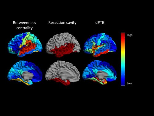
Epilepsy surgery is a potent treatment for refractory epilepsy, however, one-third of patients continue to experience seizures after surgery. Our aim was to develop a new method to identify the epileptogenic zone pre-operatively based on network theory. We analyzed eyes-closed resting-state MEG recordings of 22 patients with refractory epilepsy. Beamformer-based time-series were reconstructed for 90 AAL atlas regions. We analysed 20 epochs of 3.28s without artefacts or epileptiform activity. We calculated betweenness centrality (an indicator of hubs) based on the phase lag index (PLI) and the minimum spanning tree. Furthermore, we estimated directed phase transfer entropy (dPTE) (an indicator of information senders) for different frequency bands. ROIs with high broadband betweenness centrality (hubs) coincided with the resection cavity (or resection lobe) in 8/14 (9/14) seizure-free patients and in 0/8 (0/8) patients with remaining seizures (73% (77%) accuracy). High dPTE values coincided with the resection cavity (or resection lobe) in 8/14 (10/14) seizure-free patients and only in 2/8 (2/8) patients with remaining seizures (64% (73%) accuracy) in the delta band (0.5-4Hz). Hub regions and strong senders are markers of the epileptogenic zone. This is a first step towards a method that can be applied to MEG recordings even without epileptiform activity.
Figure: The resection cavity (red, middle column) coincided with hub regions (high betweenness centrality, left column) and strong sender regions (high dPTE, right column) in this seizure-free patient. |

