Introduction
The aim of the Cognomics project is to mimic in vivo neuronal circuits in vitro. The study concerns cognitive processes associated to reflex, pain and learning circuits and is based on the culture of dissociated neuronal cells extracted from rats. Additionally, the existence of a relationship between the results obtained from cultured neuronal circuits with the ones resulting from cognitive studies in humans will be investigated. The knowledge on the dynamics and coding of activity and learning obtained from these artificial networks will be useful, for example, to improve the process of training cerebrovascular accident patients with rehabilitation robots, as well as to gain new insights in the physiology and pathophysiology of the nervous system.
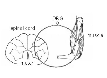
Figure 1 Schematics of the spinal reflex circuit, one of the processes to be mimicked. The system is composed by muscle spindles, dorsal root ganglion (DRG) cells, motor neurons in the ventral horn of the spinal cord and the effector muscle.
This project is carried out in collaboration with the Biomedical Signal and Systems (BSS), Biomedical Mechanical Engineering (BW) groups, as well a the Neurosurgery dept. of the LUMC. The main task of the BIOS group in the framework of this project is to design and fabricate chips that will serve as platforms for the culture of neuronal cells containing the necessary elements to mimic the previously mentioned cognitive processes. The challenge is the combination of different elements and techniques for the fabrication of the chips. Micromachining (microlithography, reactive ion etching and wafer bonding) will be employed to define culture chambers, microfluidic and interconnecting channels. Microfluidic channels are necessary for adding and subtracting substances such as nutrients and drugs. Electrical stimulation and registration of neuronal activity will be possible by means of micro electrode arrays (MEA) and the incorporation of ISFETs (ion sensitive field effect transistors) will make it possible to determine the concentration of ions. The combination of MEAs and microfluidic channels is called micro-Channel Electrode Arrays (mCEA’s).
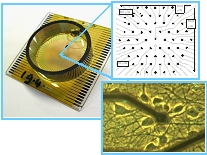
Figure 2. Standard microelectrode array showing (upper photograph) 62 electrodes and (lower photograph) one electrode covered with neurons. The planned device will contain a network of culture chambers and microfluidic channels.
Interested?
If you are interested and for instance would like to do your graduation work or practical term, please contact via the email address below.
Contact Information
Eddy de Weerd and / or Wouter Olthuis
MESA+ Institute for Nanotechnology
University of Twente
P.O. Box 217
7500 AE Enschede
The Netherlands
Phone: +31 (0)53 489 2661 (Eddy) / +31 (0)53 489 2688 (Wouter)
Fax: +31 (0)53 489 3595
Email: E.L.deWeerd@utwente.nl / W.Olthuis@utwente.nl