The focus of this theme is on the development of microfluidic systems for (bio)-medical applications, thereby increasing the knowledge of biological systems and improving the diagnostics and treatment of diseases. Microfluidics are a perfect tool for this, since dimensions comparable to single cells are used, low sample volumes are needed and multiple functionalities can be integrated in one platform. As an example electrodes can be integrated in in these devices, making electrical measurements on single cells such as sperm cells, or a tissue layer like the blood-brain barrier possible. By carefully analyzing these electrical signals, information about the morphology of the sperm cell or tightness of the tissue layer can be retrieved, which can be used to gain information about the semen quality or the functioning of the blood-brain barrier. Additional useful information can be retrieved form (automated) optical analysis using for instance fluorescence or bright field microscopy. So using these and other (new) techniques in combination with microfluidics, biomedical microdevices are developed that can finally be of use in the clinic. There are collaborations with other research groups at this and other universities, but also with doctors at the hospital, making it an applied multidisciplinary research area.
Research projects:
- The assessment and selection of spermatozoa for assisted reproductive technologies, such as IVF and ICSI (Veni grant)
- Development of a microfluidic chip platform for monitoring the monoclonal antibody concentration in blood (Saxion University, Pharmacy MST)
- A point of care sensor for cardiac biomarkers using nanofluidics separation and preconcentration
- Development of an in vitro tissue model of the blood-brain barrier (collaboration with drug delivery (UT), nanobiophysics group (UT) and LUMC)
- The creation a physiological relevant blood-brain barrier on chip system by improving the membrane (part of VESCEL project)
- Development of a microfluidic platform for early cancer diagnostics
- Measurement of biomarkers for mesenteric ischemia (collaboration with MST)

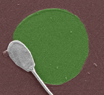
Trapping of a single spermatozoon on a protein spot.

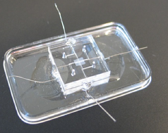
The blood-brain barrier chip.

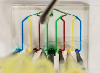
Example of a multiplex organ-on-chip device with individually addressable channels.

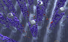
Schematic illustration of the capture of specific DNA from a sample on chemically functionalized nanopillars.
For more information contact:
