|
|
The Interventional Molecular Imaging Lab (Department of Radiology, Leiden University Medical Center) has a high multidisciplinary nature and consists of enthusiastic scientists with a background in biology, medicine, chemistry, nanotechnology, engineering and/or physics. Jointly we pursue the development of imaging agents and imaging technologies that can be applied during surgical and/or radiological interventions (image-guided surgery), ultimately aiming to improve outcome and reduce side effects. Here, we strongly focus on a multimodal/hybrid guidance concept whereby pre- and intra-operative imaging information is integrated to provide optimal sensitivity and specificity. By exploiting the different backgrounds present within the group, we approach (clinical) challenges from multiple angles. When combined, this multidisciplinary approach can drive a rapid clinical translation of new technologies that could benefit patient care.
BIOGRAPHY
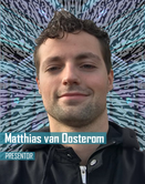
RESEARCHER & ENGINEER Matthias is both a researcher and an engineer who has pursued the development and translation of several novel image-guided surgery techniques for the operating room. After obtaining his MSc degree in Applied Physics (Delft University of Technology), he worked as a PhD (Department of Surgery and Radiology, Leiden University Medical Center) with an aim to advance precision surgery. To this end he worked on and published on a number of medical devices for robotic surgery, radio guided surgery, fluorescence-guided surgery and surgical navigation. He now holds positions as a post-doctoral researcher with the Interventional Molecular Imaging Laboratory (Department of Radiology, Leiden University Medical Center) where he heads the engineering projects of the group, as well as guest researcher at the Department of Urology of the Netherlands Cancer Institute – Antoni van Leeuwenhoek Hospital.
PRESENTATION
Robot-assisted laparoscopic surgery is slowly becoming the new standard for minimal-invasive laparoscopic procedures. In the pursue of precision surgery, however, precise instruments should be complemented with accurate definition of the surgical targets. To this end, image-guided surgery could provide an ideal complementing companion for the robotic platform. An important aspect within image-guided surgery is the development of tracers that target specific anatomies or diseases in the patient. In addition, another often overlooked aspect is the integration of the imaging and detection modalities in the surgical workflow, something particularly relevant in the complex robotic setup.
Current surgical robotic platforms are often equipped with a fluorescence imaging laparoscope, while use of multiple fluorescent tracers is explored to differentiate between different anatomical structures. For the application of radioguidance, the traditional laparoscopic gamma probes have proven cumbersome in the robotic setting. Therefore, tethered DROP-IN gamma and beta probes have shown great promise for both the sentinel lymph node and PSMA-targeted prostate cancer procedures. Hybrid approaches, providing both radio- and fluorescence-guidance, have shown to provide complementary guidance at specific stages of the surgical procedure. Finally, surgical navigation of the imaging modalities based on 3D patient scans (e.g. SPECT/CT) has been suggested to optimally connect pre- and intraoperative imaging modalities.
From a technical perspective, many engineering advances are now being developed for image-guided robotic surgery, working towards precise, less invasive and more effective surgical procedures.


