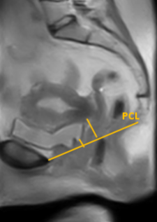
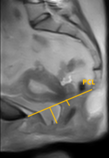
Pelvic organ prolapse (POP) is a common condition in women above 40 years. Symptoms can either be relieved with a pessary or, when this is unsuccessful, with surgery. However, pessaries generally fail over time and the percentage of recurrence after surgery is high. Nowadays, MRI is only performed in supine position for complex POP cases. We have shown that the supine position is inconsistent with the maximal extent of POP using our low-field MRI system where the patient can be scanned in upright position.
Our main question in this research is: “Can we improve the quality life and/or reduce the amount of re-interventions for women who suffer from POP by gaining additional anatomical information from MRI scans?”. The goal of this research is to first get a better understanding of the underlying anatomy and function of the pelvic floor by imaging in a posture that coincides with the complaints (i.e. upright). Therefore this part of the research mainly focusses on the low-field rotating MRI scanner. Subsequently, the effect of the treatment options, both pessary and surgery, will be evaluated and analyzed. Since this might require higher quality than what is possible on the low-field scanner, this can be performed on the 1.5T scanner. Imaging of muscles in 3D and muscle damage and function is of interest here.


The following two examples of a mid-sagittal view show the clear influence of gravity on the position of the pelvic organs. The left image was made in supine position and the right image in upright positon
This research is performed in collaboration with dr. Anique Bellos-Grob, post-doc in the M3I group, and is the main focus of the PhD project of Lisan Morsinkhof. Outside of the UT we have cooperation from the Gynaecology departments of the UMC Utrecht, Medisch Spectrum Twente, Ziekenhuisgroep Twente and the Radboud UMC.
