High-Resolution Cryo-Electron Tomography – Current State and Future Prospects
Sebastian Tacke and Stefan Raunser
Department of Structural Biochemistry, Max Planck Institute of Molecular Physiology, Dortmund, Germany
Electron tomography at cryogenic temperatures (cryo-ET) offers the possibility to structurally characterize biological macromolecules and macromolecular assemblies in their native environment. By using sub-tomogram averaging, three-dimensional reconstructions at near-atomic resolution can be obtained [1]. A typical workflow of current cryo-ET approaches is illustrated below.
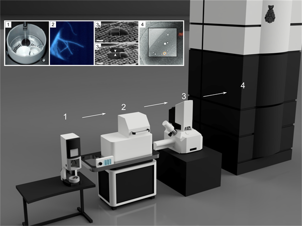
In the first step, the sample is vitrified in liquid ethane. This guarantees that the structure of the biological sample is preserved [figure 1.1, 2, 3]. In case the region of interest is difficult to identify, cryo-light microscopy is applied [figure 1.2, 4]. Since many biological samples are too thick to be penetrated by the electron beam, the sample material is then thinned by a focused ion beam [figure 1.3, 5]. Finally, cryo-ET tilt series are acquired of the thinned regions, called lamellae [figure 1.4, 6].
The entire workflow involves numerous handling steps, most of which under cryogenic conditions and in an anhydrous environment to avoid de-vitrification or contamination of the sample material. Due to the enormous complexity, only a small number of labs have so far established this technique worldwide.
Here, I will describe the current state-of-the-art workflow, and discuss open issues and necessary improvements to make cryo-ET accessible for many users in the near future.
[1] F. K. M. Schur et al. (2016) An atomic model of HIV-1 capsid-SP1 Reveals structures regulating assembly and maturation. Science. 353, 506-508.
[2] P. Echlin. Low-Temperature Microscopy and Analysis. Plenum Publishing Corporation, New York, 1992.
[3] P. Zhang. (2013). Correlative cryo-electron tomography and optical microscopy of cells. Current Opinion in Structural Biology. 23, 763-770.
[4] R. Koning et al. (2014). Correlative cryo-fluorescence light microscopy and cryo-electron tomography of Streptomyces. Methods in Cell Biology. 124, 217-239.
[5] A. Rigort et al. (2012). Focused ion beam micromachining of eukaryotic cells for cryoelectron tomography. PNAS. 109, 4449-4454.
[6] Y.-W. Chang et al. (2014). Correlated cryogenic photoactivated localization microscopy and cryo-electron tomography. Nature Methods. 11, 737-739.