INSTRUCTIVE 3D MEMBRANES FOR A PHYSIOLOGICALLY INSPIRED SYNOVIUM-ON-CHIP MODEL
Background: The synovium or synovial membrane contributes to the production of synovial fluid, which not only supplies nutrients to the articular cartilage but also lessens articulation related friction. The synovial membrane is composed by two layers: a thin and highly cellular lining layer composed of two cell types: synovial fibroblasts and synovial macrophages, and a supporting layer of loose connective tissue containing fibroblasts, nerves, blood and lymphatic vasculature1. Besides producing extracellular matrix, the synovial fibroblasts, secrete hyaluronic acid and lubricin into the synovial fluid. Synovial tissue macrophages (STM) are constitutively resident in healthy synovium. The different macrophage subpopulations in the human synovium have the potential to contribute either to joint homeostasis or to chronic inflammation, such as rheumatoid arthritis (RA)2. Current in vitro models lack in complexity and dynamic cues3, thus limiting our knowledge regarding their relative involvement in the articular joint homeostasis. State-of-the-art organ-on-chip technology provides a promising platform to recreate physiologically relevant 3D micro-environments.
We have previously developed complex instructive membranes based on electrospinning and 3D melt-writing, which allows us to closely mimic the native synovial membrane. In this context, further research using this model will include the incorporation and characterization of these 3D membranes on-chip, to study the migratory and inflammatory patterns of synovium cells, e.g. STM. We hypothesise that the synovium-on-chip model will contribute to further understanding the role of human STM in the regulation of joint homeostasis, which may accelerate the development of new therapeutic strategies for RA.
Goal: Contribute to yielding insights into the migratory and inflammatory patterns of synovium cells, using a microfabricated synovium-on-chip platform.
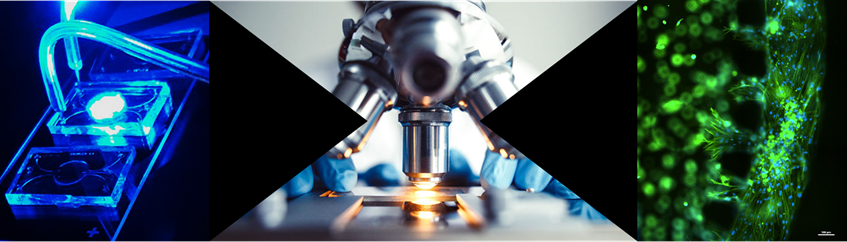
Techniques applied:
- Micro-fabrication
- Cell culture
- Biochemical assays
- Microscopy
Relevant literature:
1 Kinne, R. W., Stuhlmüller, B. & Burmester, G.-R. Cells of the synovium in
rheumatoid arthritis. Macrophages. Arthritis Research & Therapy 9, 224,
doi:10.1186/ar2333 (2007).
2 Kurowska-Stolarska, M. & Alivernini, S. Synovial tissue macrophages:
friend or foe? RMD open 3, e000527-e000527,
doi:10.1136/rmdopen-2017-000527 (2017).
3 Stefani, R. M. et al. A Functional Tissue-Engineered Synovium Model
to Study Osteoarthritis Progression and Treatment. Tissue Engineering Part A,
doi:10.1089/ten.tea.2018.0142 (2018).
More information at: