Staging osteoarthritis by 2-photon second harmonic generation microscopy
This project is suitable for BMT students with affinity for technology, and APh students with affinity for biology. Ideally, we would like to have a BMT student and an APh student that work simultaneously (and together) on the different aspects of this project.
Background:
Cartilage is a tissue with low self-repair capability and therefore it is important to find therapeutic strategies that enables efficient cartilage repair. Osteoarthritis (OA) is a gradual process, characterized by loss of structure, loss of cartilage specific proteins such as glycosaminoglycans and collagen 2, and increase in hypertrophic markers such as collagen 10 (see figure). Currently, the severity of OA is determined either by X-ray of the joint space narrowing1, macroscopically using a grading system devised by Collins in the 1940’s 2, or histologically using the OARSI scoring3 as used for staging the cartilage in the figure below. Although quite well described the OARSI grading is still subjective, and therefore multiple assessors are needed to score the cartilage.
At the molecular level, cartilage degeneration is characterized by a loss of glycosaminoglycans and collagen 2. In addition, the alignment of collagen fibrils is lost in OA. We hypothesize that both the decrease of collagen content and the loss of the collagen structure can be visualized and objectively quantified using 2-photon second harmonic generation (SHG) microscopy 4.
Aim: In this MSc project, we would like to test the hypothesis on cartilage explants that have previously been isolated and of which we have thin 5 mm slices.
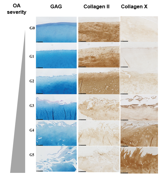
Figure 1: Alcian blue staining of Glycosaminoglycans (GAG), and immunohistochemistry of Collagen 2 and Collagen 10 in human cartilage explants with increasing osteoarthritis from healthy (G0) to endstage (G5) OA. These (immuno)histological stainings can be used to grade the cartilage degeneration.
Techniques for both BMT and APh students:
- Imaging of thin slices of cartilage explants that have been previously made
- Data analysis, e.g. determine volume collagen in total volume sample
- Polarisation imaging using Picrosirius Red (now standard in Cartilage Tissue Engineering).
Specific for BMT student:
- Isolation and slicing of cartilage explants from cow
- Histological staining for OARSI scoring of damaged cartilage
Specific for APh student:
- Adjusting the microscope set-up: e.g. polarisation module
- Advanced image analysis, e.g. quantifying tissue heterogeneity and disorder, tracing collagen fibre directions through 3-D image construction, machine-learning approaches to replace OARSI scoring.
References:
1 Hayashi, D., Roemer, F. W., Jarraya, M. & Guermazi, A. Imaging in
Osteoarthritis. Radiol Clin North Am 55, 1085-1102,
doi:10.1016/j.rcl.2017.04.012 (2017).
2 Collins, D. H. Osteoarthritis. 74-115 (Edward Arnold & Co, 1949).
3 Pritzker, K. P. H. et al. Osteoarthritis cartilage histopathology: grading and
staging. Osteoarthr Cartilage 14, 13-29, doi:10.1016/j.joca.2005.07.014.
4 Chaudhary, R. et al. Articular cartilage zonal differentiation via 3D
Second-Harmonic Generation imaging microscopy. Connective tissue
research 56, 76-86, doi:10.3109/03008207.2015.1013192 (2015).