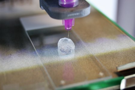
For some types of cancer, it is the human body itself that complicates adequate treatment. Our connective tissue and immune system then build a sort of ‘defensive wall’ around the tumor, through which chemo therapy can hardly find its way. University of Twente researcher Marcel Heinrich mimicked this tumor micro environment in the lab using 3D modelling and bioprinting. If lab research enable us to actually find new ways of reaching the tumor, it is also possible to reduce the number of tests on animals. Heinrich recently finished his PhD research cum laude.
Pancreatic cancer and glioblastoma – an aggressive brain tumor – are two types of cancer with a bad prognosis. One of the reasons for this is, that it is very difficult to intervene. This is caused by the micro environment that the body’s immune system and connective tissue uses. Chemotherapy has been designed to destroy the tumor, but now hits on an environment that makes access to the tumor very difficult. And: the pancreatic cancer environment is totally different from the one around glioblastoma. Two dimensional (2D) fail to describe this sufficiently well, that is why Marcel Heinrich modelled the environments in 3D. He also used bio printing techniques to print the environment, with patient data as the input. The basic question is: what is needed for opening up this ‘defence wall’ and give chemo treatment the necessary access?
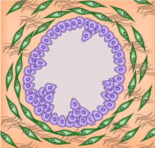
The typical tumor environment of pancreatic cancer doesn't give easy access to treatment. Tumor cells are purple, fibrotic stroma green.
Different strategies
 There is no ‘one size fits all’ solution for a tumor environment. Around the pancreatic cancer (PDAC: pancreatic ductal adenocarcinoma) a strong network of fibrous tissue builds up, fibrotic stroma, that can obstruct the well-functioning of blood vessels. A glioblastoma attracts macrophages, a type of white blood cells that in this case ‘help the tumor’. It may be clear that for each type, a different access strategy is needed. The 3D-models of the tumor environment help reaching this goal by experimenting based on data that is trustworthy. Unlike in tissue engineering, the printed 'mini pancreas' and 'mini brain' aren’t created for implant. They just have a minimum of basic functions, characteristic for the tumor, needed for evaluating strategies to reach it for treatment. As the models give the researcher a highly valuable level of freedom to do experiments without using animals, the amount of animal tests can be reduced drastically, says Marcel Heinrich.
Marcel Heinrich (Herne, Duitsland, 1992) did his research in the Advanced organ bioengineering and therapeutics group, part of University of Twente’s TechMed Centre. He recently defended his PhD thesis ‘Engineering the tumor microenvironment – novel 3D in vitro models to study cellular interactions & therapeutics’. His supervisor was Prof Jai Prakash.


