


Over the last two decades, nuclear medicine has gained tremendous attention and is nowadays a key component in the clinical management of patients. Nuclear imaging techniques, such as positron emission tomography (PET), have become clinical routine and is superior as a diagnostic tool mainly in the field of medical oncology, cardiology and neurology.

Figure 1. Molecular imaging technique used to measure in vivo processes.
PET Imaging
PET imaging is a molecular imaging technique used to measure in vivo processes noninvasively. This technique is often combined with magnetic resonance imaging (MRI) or computed tomography (CT) to provide better anatomical information. In PET radioactive isotopes are introduced into biologically active molecules which are injected at tracer (subpharmacological) doses into the body. Radionuclides used in PET scanning are typically isotopes with short half-lives such as 11C (~20 min), 15O (~2 min), 18F (~110 min) and 68Ga (~68 min). Unstable radioactive atoms decay by positron emission to become stable. The decay of radionuclides occurs by the emission of a positively charged particle called positron (β+). The positron passes a short distance (1–5 mm), dependent on the positron energy of the isotope, in the surrounding tissue before it annihilates by combining with an electron. Then, the mass of the positron and electron is converted to energy resulting in two 511 keV γ-rays which are emitted simultaneously at approximately 180° to each other. The pair of γ-rays is detected by rings of detectors in the PET scanner. The acquisition of a large number of coincident events provides data which can be reconstructed to an image with information on the spatial distribution of radioactivity as a function of time.
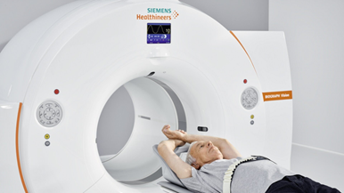
Figure 2. Clinical PET scanner
Facilities
State of the art facilities are available at UMCG to develop tracers, implementation and GMP production of tracers and evaluation of tracers. UMCG is equipped 18MeV twin proton cyclotron (IBA), GMP laboratory facilities, clinical and pre-clinical hybrid scanners (Siemens). The department Nuclear medicine & molecular imaging (NMMI) has a strong national and international reputation in PET methodology along the whole axis of radionuclide production, radiochemistry, clinical physics and clinical PET implementation. With respect to radiochemistry, a unique international position has been reached regarding tracer development and tracer production for clinical research, with appropriate EU GMP production licenses.
The NMMI department is embedded in research institutes for both oncology, cardiology and neurosciences. Intensive collaborations exist with national and international biotechnology and pharmaceutical industries with regard to the development of new drugs, and with imaging industries with regard to innovation of molecular imaging technology.
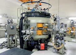
Figure 3. Cyclotron for the production of radionuclides for Imaging, like 18F, 11C, 13N and 15O.
Tracers
At the UMCG-GMP production facility, a broad assortment of tracers is available for clinical care and clinical research. At this moment more than 25 different tracers are available. Each specific tracers reflects a pathological or biological process. Tracers are available for in vivo measurement of perfusion, glucose metabolism, inflammation, amino acid uptake in tumours. Amongst others, tracers like 18FDOPA, 18FES are available for measuring several (brain) receptors and transporters.
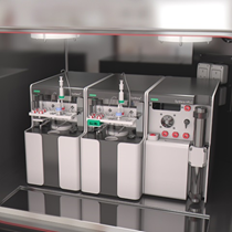
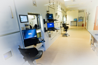
Figure 4. GMP production facility (right) equipped with GMP compliant synthesis modules.
Contact